
Question
A 40 year old motorcyclist is hypotensive in the emergency department after a high speed crash.
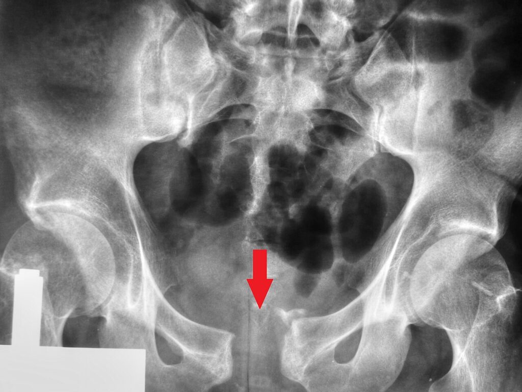
- Describe the radiograph and classify the injury.
- Outline your initial management of the haemodynamically unstable patient.
- What is the definitive management?
Answer
Radiograph
- Symphysis pubis diastasis
- Anteroposterior compression injury
- Over 2.5 cm diastasis implies sacrospinous and anterior sacroiliac ligament injury (APC II)
Young and Burgess APC types
| Type | Features |
|---|---|
| APC I | Symphysis under 2.5 cm, posterior ligaments intact |
| APC II | Symphysis over 2.5 cm, anterior SI ligaments torn, posterior intact |
| APC III | Complete SI disruption, vertically stable, highest transfusion need |
Initial management
- ATLS, massive transfusion protocol and tranexamic acid
- Pelvic binder at the level of the greater trochanters
- Exclude other sources of bleeding with FAST or CT
- Venous bleeding most common, arterial in about 15%
- Preperitoneal packing or angioembolisation for ongoing bleeding
- Anterior external fixator as temporary stabilisation
- Examine perineum, rectum and vagina for open injury, check for urethral injury before catheterisation
Definitive
- Symphyseal plating through a Pfannenstiel approach
- Iliosacral screws for posterior SI disruption in APC III
- Thromboprophylaxis
Related pages
Author Contributions
Orthofracs team
Reviewed by Professor Phong Tran, Head of Orthopaedic Surgery, Western Health. Last updated 10 October 2026.