
Question
A 34 year old man falls from a scaffold. He has right hip pain with an intact sciatic nerve.
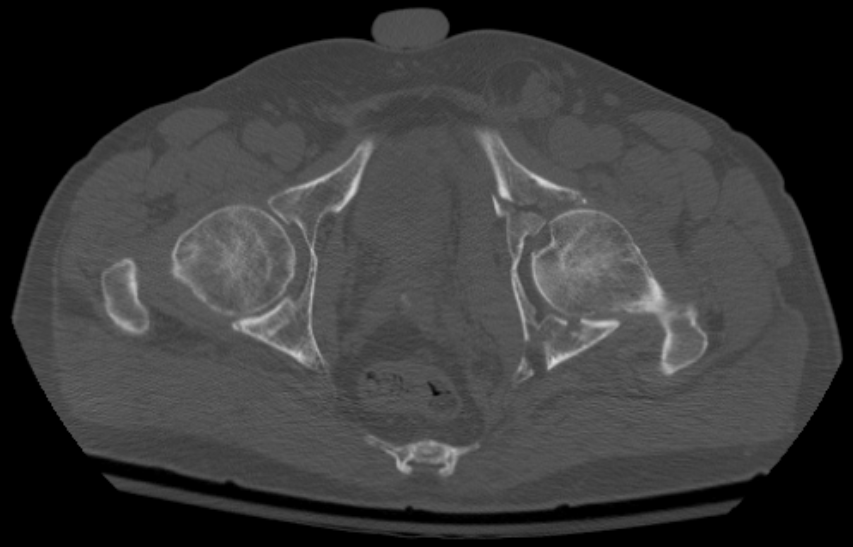
- What radiographic views and lines do you assess?
- Classify acetabular fractures.
- What are the indications for surgery and the approaches?
Answer
Views and lines
- AP pelvis, Judet obturator oblique and iliac oblique views
- Iliopectineal line (anterior column)
- Ilioischial line (posterior column)
- Anterior and posterior walls
- Teardrop and roof
- CT with 3D reconstruction
Letournel classification
| Elementary | Associated |
|---|---|
| Posterior wall | Posterior column and posterior wall |
| Posterior column | Transverse and posterior wall |
| Anterior wall | T shaped |
| Anterior column | Anterior column and posterior hemitransverse |
| Transverse | Both column |
Indications for surgery
- Articular displacement over 2 mm in the dome
- Roof arc angles under 45 degrees
- Posterior wall fragment over 40 to 50% or instability
- Incarcerated fragments
- Non concentric reduction
Approaches
- Kocher-Langenbeck for posterior wall and column
- Ilioinguinal or anterior intrapelvic (Stoppa) for anterior column
- Combined or extended iliofemoral for complex patterns
- Acute THA for elderly patients with dome impaction or femoral head damage
Related pages
Author Contributions
Orthofracs team
Reviewed by Professor Phong Tran, Head of Orthopaedic Surgery, Western Health. Last updated 10 October 2026.