
Question
A 74 year old woman had an intertrochanteric fracture fixed with a cephalomedullary nail. Eight weeks later she has increasing hip pain.
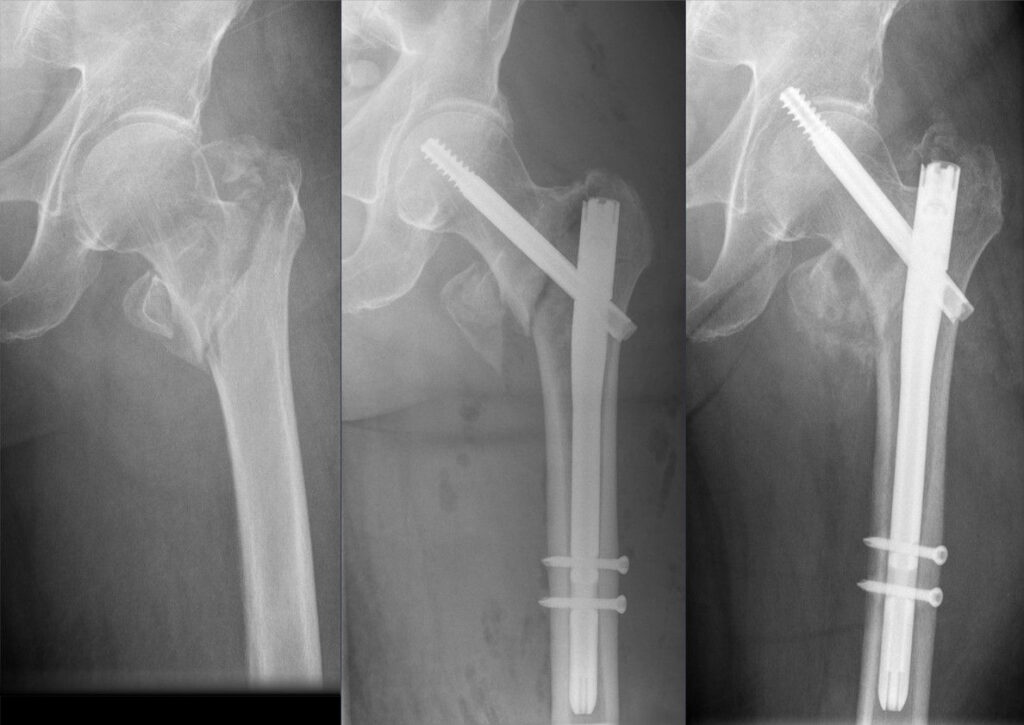
- Describe the series of radiographs.
- What are the risk factors for this complication?
- How would you manage it?
Answer
Radiographs
- Intertrochanteric fracture fixed with a cephalomedullary nail
- Later films show varus collapse and the lag screw cutting out of the femoral head superiorly
Risk factors
- Tip apex distance over 25 mm (Baumgaertner)
- Superior or anterior lag screw position
- Varus or poor reduction
- Unstable fracture pattern
- Poor bone quality
- Calcar referenced tip apex distance adds information
Management
- Exclude infection with bloods and aspiration or tissue samples
- Assess acetabular damage from the screw
- Conversion to total hip arthroplasty in most elderly patients
- Calcar replacing or long stem, with trochanteric fixation as needed
- Revision fixation with valgus reduction in younger patients with good head stock
- Higher dislocation and complication rates than primary THA
Related pages
Author Contributions
Orthofracs team
Reviewed by Professor Phong Tran, Head of Orthopaedic Surgery, Western Health. Last updated 10 October 2026.