
Question
A 42 year old man has a year of mechanical hip pain with intermittent catching.
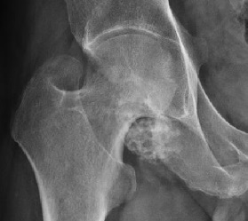
- Describe the radiograph and give the diagnosis.
- What are the pathology and differential diagnosis?
- How is it treated?
Answer
Radiograph
- Multiple small, rounded, calcified loose bodies around the hip
- Similar size and shape
- Possible extrinsic erosion of the femoral neck
- Primary synovial chondromatosis
Pathology
- Synovial metaplasia producing cartilaginous nodules
- Nodules detach and calcify, may enlarge in synovial fluid
- Recurrent FN1 and ACVR2A gene fusions suggest a neoplastic process
- Milgram stages, intrasynovial active, transitional, multiple loose bodies with quiescent synovium
- Malignant change to chondrosarcoma is rare (under 5%)
Differential diagnosis
- Secondary osteochondromatosis from osteoarthritis (fewer, varied loose bodies)
- Pigmented villonodular synovitis
- Synovial chondrosarcoma
Treatment
- Loose body removal and synovectomy
- Arthroscopic or open with surgical dislocation
- Recurrence 10 to 20%
- THA for established arthritis
Related pages
Author Contributions
Orthofracs team
Reviewed by Professor Phong Tran, Head of Orthopaedic Surgery, Western Health. Last updated 10 October 2026.