
Question
A 79 year old man falls and has a shortened, externally rotated right leg.
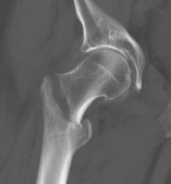
- Describe the imaging.
- What features make this fracture unstable?
- Discuss your choice of implant and how to avoid failure.
Answer
Imaging
- Pertrochanteric (intertrochanteric) fracture of the proximal femur
- Assess posteromedial comminution, lesser trochanter, lateral wall and reverse obliquity
- Full length femur radiographs before nailing
Unstable features
- Posteromedial comminution with a large lesser trochanter fragment
- Thin or fractured lateral wall (under 20.5 mm)
- Reverse obliquity
- Subtrochanteric extension
- AO 31A2 and A3 patterns
Implant choice
- Sliding hip screw for stable A1 patterns
- Cephalomedullary nail for unstable, reverse oblique and subtrochanteric extension
- Reduce on a traction table before fixation
- Lag screw centre centre or inferior on AP, centre on lateral
- Tip apex distance under 25 mm (Baumgaertner)
- Avoid varus reduction
- Weight bear as tolerated
Failure
- Cut out of the lag screw, the most common mechanical failure
- Excess collapse and medialisation
- Peri-implant fracture
Related pages
Author Contributions
Orthofracs team
Reviewed by Professor Phong Tran, Head of Orthopaedic Surgery, Western Health. Last updated 10 October 2026.