
Definition
- Primary malignant neoplasm of bone & or soft tissue which contains fibrous & histiocytic elements
Epidemiology
- Rare
- 1 per million or one per 2 million
- Affects older age group 40-70
- More common in males
Site
- Metaphysis of long bones, more so lower limbs than upper limbs
Aetiology
- 20% arise secondary to other lesions such as fibrous dysplasia, bone infarction, Paget’s disease & irradiation
Pathology
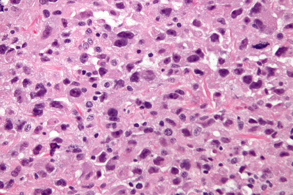
- “storiform pattern”
- there is an appearance of wheel spokes radiating from a slit like vessel, in a fibrous background
- A storiform pattern is not however diagnostic of MFH because it is also seen in osteosarcomas, leiomyosarcomas, neurogenic sarcomas, haemangiopericytomas & non ossifying fibromas
- Other typical histological features are bizarre histiocytic cells, numerous mitotic figures, & scattering of chronic inflammatory cells
Histological variants:
- MFH – myxoid subtype
- Sheets of myxomatous tissue with large histiocytes & abnormal mitoses
- MFH – Inflammatory subtype
- Field of malignant histiocytes peppered with acute inflammatory cells, which may obscure the fibrous elements
- MFH – Giant cell type
- Large number of multinucleated giant cells
- Stout’s hypothesis – theory of histiocytic & fibrous origin: histiocytes are derived from bone marrow monocytes – “tissue macrophages”. Histiocytes have the potential to transform to spindle cells which produce collagen, & are indistinguishable from fibrocytes
- MFH is a diagnosis of exclusion; if the tumour resembles MFH but produces regions resembling another malignant tumour it is diagnosed as that tumour
Clinical
- Pain, swelling, pathological fracture
- Relatively high metastatic potential, usually to lungs & other bones
Investigations
Laboratory
No characteristic alterations
X-ray
- Lesions usually lytic or permeative; may expand cortex without breaching it
- Frequent periosteal reaction
- High index of suspicion if permeative radiolucency adjacent to bone infarct, fibrous dysplasia or Paget’s disease
MRI
- T1 – low or intermediate intensity
- T2 – high intensity
Immunohistochemistry
- Vimentin stain positive
- More specific is A1-AT antibody which also binds normal histiocytes
Treatment
- Induction chemotherapy
- Limb sparing surgery
Prognosis
- Poor – around 60% 5ysr, but probably has a better prognosis than other sarcomatous lesions
- Prognosis worse for secondary lesions. 40% alive at 4 years
Reviewed by Professor Phong Tran, Head of Orthopaedic Surgery, Western Health. Last updated 10 October 2026.