
ACJ Arthritis
Summary
- Osteoarthritis of the Acromioclavicular joint
- The AC joint can become a source of pain in the shoulder, because of:
- degenerative osteoarthritis,
- posttraumatic arthritis,
- distal clavicle osteolysis.
- Diagnosis
- Pain on palpation,
- reproduction of pain with cross-body adduction
- diagnostic injections
- Plain radiographs
- Zanca view,
- Treatment
- Nonoperative management
- can be successful,
- Operative
- distal clavicle resection
- Nonoperative management
Aetiology
- idiopathic
- elderly
- Degenerative osteoarthritis is more common with advanced age
- AC joint arthritis is much less common than hip, knee, or glenohumeral arthritis
- elderly
- trauma
- young
- Posttraumatic arthritis is a more common cause than primary osteoarthritis
- young
- Distal clavicle osteolysis
- less-common cause, occurs in certain power athletes (e.g., weight-lifters).
Epidemiology
- Common cause of superior shoulder pain in middle aged and older adults
- Radiographic degeneration very common after 40 years, often asymptomatic
- Post traumatic arthritis after ACJ injury or distal clavicle fracture
- Distal clavicle osteolysis in young weightlifters
- Coexists with subacromial impingement and rotator cuff disease
Anatomy
- The AC joint is a hyaline cartilage joint with a fibrocartilage meniscal disc.
Pathology
- Disc starts to break down with normal ageing and, by early adulthood, is minimal
- Disc may be injured with an AC separation or with repetitive activity
Classification
4 patterns
| Pattern | Notes |
|---|---|
| DJD with osteophytes | Contribute to impingement |
| Osteolysis | With resorption & gross osteoporosis, due to repetitive microtrauma (eg weight lifters) |
| RA | |
| Hyperparathyroidism |
History
- Pain:
- anterosuperior shoulder pain
- worse with activities that load the joint (reaching across shoulder, behind body, bench-press)
- Night pain: difficulty sleeping on affected side
- pain radiates to trapezius > spasm
Examination
- Direct Palpation
- Tenderness to direct palpation
- is most reliable sign
- may feel osteophytes
- Tenderness to direct palpation
- Provocative tests
- horizontal adduction of arm (tends to overlap with impingement)
- max IR of shoulder (more sensitive & specific)
- Injection of local anaesthetic
- useful
Investigations
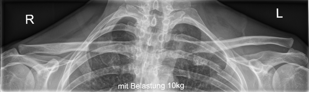
X-ray
- AP Shoulder
- neutral
- IR
- ER (true AP)
- AP with 10° cephalic tilt with 1/3 penetration [zanca view]
- best for AC joint
- Look for
- sclerosis
- subchondral cysts
- joint space narrowing
- osteophyte formation
- bone loss at distal clavicle (osteolysis)
Bone Scan
- increased uptake in joint
MRI
- very helpful
- can be difficult to determine signal because of Normal age related changes
Lignocaine Injection
- Local anaesthetic injected into the ACJ, often with corticosteroid
- Pain relief confirms the ACJ as the pain source
- Palpation guided injection often misses the joint, US guidance improves accuracy
- Separate subacromial injection helps distinguish coexisting pathology
- Positive response predicts good outcome from distal clavicle excision
Differential Diagnosis
- Intrinsic
- Rotator Cuff Impingement
- Calcific tendinitis
- Frozen shoulder
- Glenohumeral arthritis
- ACJ gout
- ACJ sepsis
- Extrinsic
- Cervical root C4/5
- Shoulder tip pain from abdominal pathology
Treatment
Principles
- Most can be treated nonoperatively
Non-operative
- most respond to Non-operative management
- NSAIDs
- Activity modification
- Limitation of exacerbating activities such as bench-presses, dips, push-ups, and overhead activities
- Steroid injection
- Physiotherapy: minimal role
- consider operative management after 6 months
Operative
- Indication
- if fail nonoperative management
- 90% success rate
- resect 1cm – 2cm (to prevent continued bony contact between the clavicle and acromion)
- Options
- Open resection
- Arthroscopic
- preserves AC capsule
- quicker rehabilitation
- Complications
- incomplete resection
- continued pain after surgery
- excessive resection leading to distal clavicle instability
Prognosis
- Most respond to activity modification, analgesia and injection
- Good to excellent results in most patients after distal clavicle excision
- Open and arthroscopic excision give comparable results, arthroscopic recovery is quicker
- Over resection or AC ligament injury causes instability and residual pain
- Poorer results in post traumatic cases and with coexisting pathology
Reviewed by Professor Phong Tran, Head of Orthopaedic Surgery, Western Health. Last updated 10 October 2026.