
Osteonecrosis of the femoral head is death of subchondral bone from loss of blood supply, linked to trauma, corticosteroids, alcohol and sickle cell disease. It is staged with Ficat and Arlet or Steinberg. Core decompression may help before collapse, and hip replacement is needed after collapse.
Western Health Orthopaedic Registrar presentation – Osteonecrosis of the femoral head by Dr Devinder Garewal
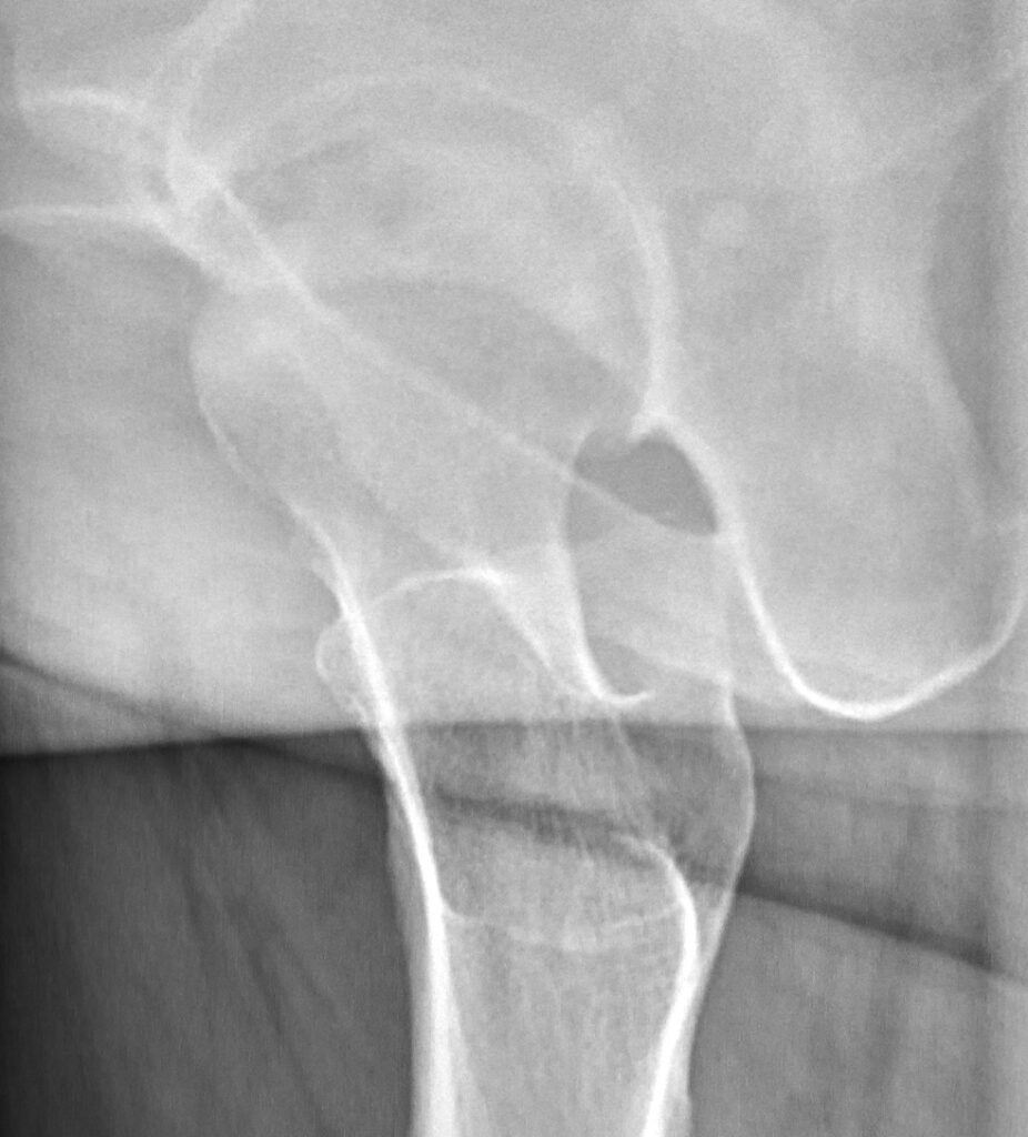
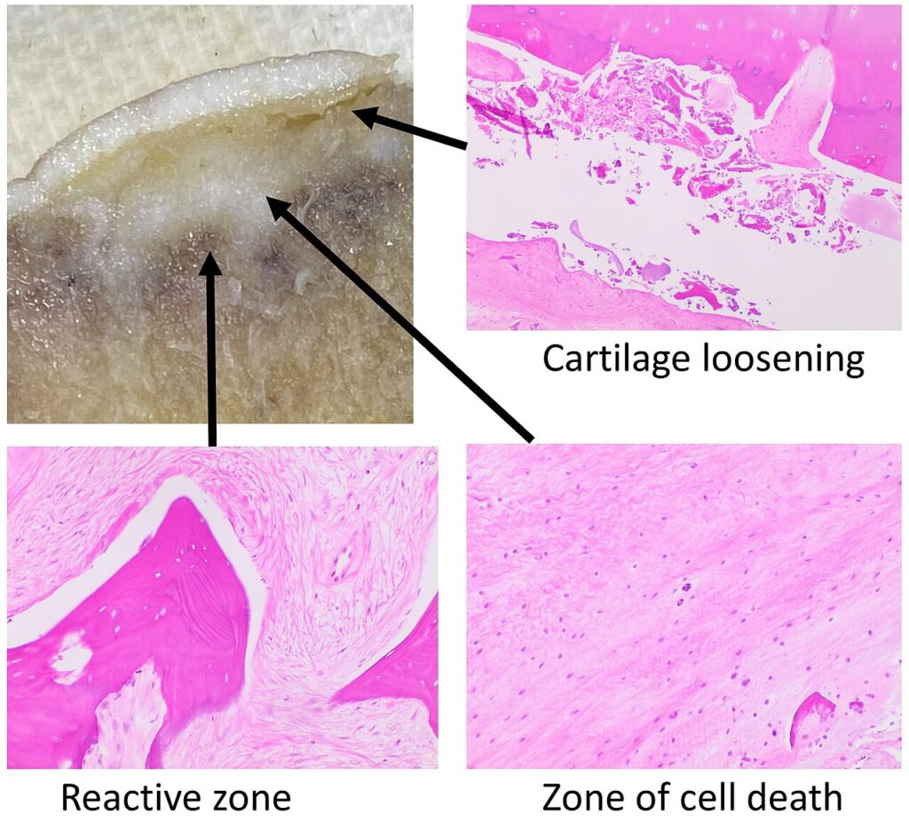
Classification
Ficat Classification
- Professor RP Ficat; Ficat Classification of Osteonecrosis of the Hip
- Normal hip with contra-lateral disease (Hungerford)
- I
- Normal radiograph
- Diagnosis following MRI, bone scan or histology
- II
- Radiographic changes of repair (osteoporosis / sclerosis / cysts)
- No osteochondral fracture
- Head spherical
- III
- Wedge shaped ↑ density
- Mottled osteoporosis
- Subchondral lucent line® Crescent sign
- Head no longer spherical “out of round”
- Usually affects antero-lateral area of femoral head (best seen on lateral view)
- IV
- Marked changes with secondary degenerative changes in the joint
- Collapse of subchondral bone & severe deformity of the head
- Steinberg Classification
- Steinberg et al JBJS (Journal of Bone & Joint Surgery) 77B: 34-41,1995
- Modified to assess extent of involvement
- More accepted classification
- Steinberg Classification of Osteonecrosis of the Hip
- Steinberg et al JBJS (Journal of Bone & Joint Surgery) 77B: 34-41,1995
| * Normal hip with contra-lateral disease (Hungerford) * Suspicion but unable to confirm | |
| I | * Normal radiograph * Diagnosis following MRI, bone scan or histology |
| II | * Radiographic changes of repair (osteoporosis / sclerosis) * No osteochondral fracture * Head spherical |
| III | * Wedge shaped ↑ density * Mottled osteoporosis * Subchondral lucent line (Crescent sign) * Head no longer spherical “out of round” * Usually affects antero-lateral area of femoral head (best seen on lateral view) |
| IV | * Flattening of the femoral head * On AP or lateral XRAYS * Now irreversible changes * Symptomatic treatment until arthroplasty |
| V | * joint narrowing with or without * acetabular involvement |
| VI | *advanced degenerative changes |
| Stage | Description |
|---|---|
| 1 + 2 | A mild: less than 15% involvement of head on XR or MRI B mod: 15- 30% C severe: more than 30% |
| 3 | A: subchondral collapse (crescent) less than 15% of artic surface B: 15- 30% C: more than 30% |
| 4 | A: less than 15% of surface has collapsed + depression is less than 2 mm B: 15- 30% collapsed or 2-4 mm depression C: more than 30% collapsed or more than 4mm depression |
| 5 | A, B or C average of femoral hd involvement as in stage 4 |
Head involvement = % of head on AP viewx % head on lateral view
- Eg 25 % involvement may be 50 % AP & 50 % lateral
Reviewed by Professor Phong Tran, Head of Orthopaedic Surgery, Western Health. Last updated 10 October 2026.