
Video
Western Health Orthopaedic Registrar presentation – Cervical Spondylosis, Radiculopathy & Myelopathy by Dr James Churchill
Definition
- Spinal cord dysfunction
- Demyelination of the lateral columns with degeneration of the anterior parts of the posterior columns
- Destruction of the central grey matter
Epidemiology
- Most common spinal cord dysfunction in patients > 55 years old
Aetiology
- Extrinsic compression of cord (or its vascular supply or both) caused by degenerative disease of spine
Pathology
- Maybe associated with congenital or developmental stenosis of the cervical canal
- Disc degenerates
- Secondary osteoarthritic changes occur in the facet & uncovertebral joints
- Reduction of available space
- Ligamentum Flavum invaginates into canal posteriorly
- Disc & posterior vertebral body osteophytes encroach anteriorly – Pincher action
Differential Diagnosis
- CVD
- AV malformation
- Demyelinating disease
- Syringomyelia
- Intracranial tumour
- Hydrocephalus
- Tabes dorsalis
- Myopathy
- Peripheral neuropathy
- Metabolic or alcoholic encephalopathy
History
- Neck pain
- Difficulty walking
- Unsteadiness on feet
- Pain, numbness & weakness with clumbsiness of hands common
- May also have radicular symptoms
- Bladder dysfunction uncommonly occurs
- co existent lumbar stenosis common
Examination
- UMN in extremities below lesion & LMN signs at level of lesion
- Can see UMN signs cephalad to lesion
- Hoffman Reflex
- Flexion of ipsilateral IPJ of index & thumb when long finger IPJ flicked & extension of neck ↑ sensitivity
- Inverted Radial Reflex
- Spontaneous flexion of digits when BR reflex elicited & indicates cord compression at C5 & C6 (commonest levels)
- L’Hermitte sign
- Flexion or extension of neck produces electric shock sensations in legs
- Abnormal Plantar reflexes
- Usually only abnormal when severe
- Earliest sensory changes are dysdiadochokinesia & poor tandem gait
- Vibration & Joint position sense affected early
- Weakness & loss of proprioception leads to Wide Based Gait
- Finger Escape Sign – deficient adduction or extension of ulnar digits of affected hand
Investigations
XRs
- Narrowed joint space
- C5/6 commonest level followed by C6/7
- Osteophytic lipping with foraminal & uncovertebral osteophytes seen
- Lateral may showListhesis
- Lordosis
- Kyphosis
- Flexion / Extension views show instability
- Pavlovs Ratio (A-P diameter of spinal canal divided by the A-P diameter of body at same level) indicator of developmental stenosis
- Should be 1.0 & < 0.8 is narrowed & stenotic
- diameter (A-P) reduced from normal (17mm) to relative (13mm) to absolute stenosis (10mm)
- Remember that 70% of population have degenerative changes by 70 year
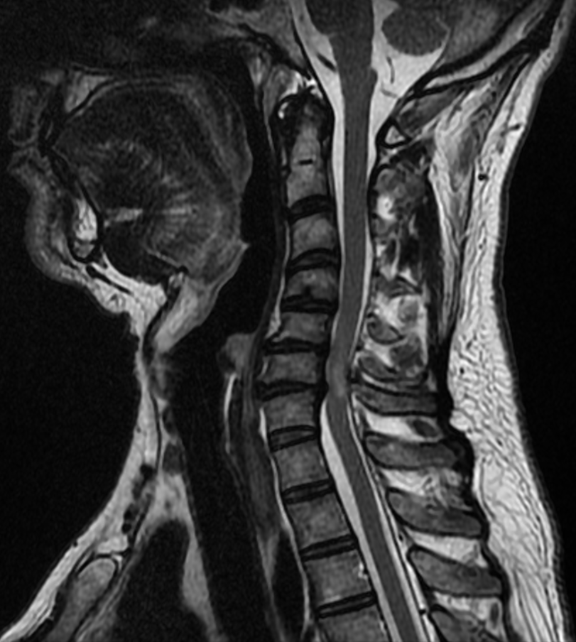
MRI
- Evaluate soft tissues of the cord
- High incidence or asymptomatic findings – 19% of asymptomatic patients have abnormality on MRI
- Disc prolapse & cord oedema with signal change seen
CT – Myelography
- Helps distinguish disc from osteophytes
- Superior for cord compression due to subluxed body & ossification of the PLL
- transverse diameter of cord at affected level on CT – Myelogram shown to be best prognostic indicator in spondylitic stenosis & ossification of PLL
Treatment
- Natural history
- suggests that > 50% of patients become worse if not treated
- best spinal cord recovery seen in those treated with:
- Decompression within 6 – 12 months
- Early, mild myelopathic signs
- Those whose transverse area of cord greater than 40 mm 2 postoperatively
Non-operative
- Cervical collar
- Nsaids
- Physiotherapy with isometric strengthening
- Ice, heat & massage
- Follow up every 6-12 weeks initially followed by yearly if no progression
- Traction & manipulation contraindicated
Operative
- Absolute Indications
- Progressive neurological deficit
- Failure to improve with 6 /12 of non op treatment
- Should try to operate prior to
- Compression ratio < 0.4
- Transverse spinal cord diam of < 40 mm2
- Increased signal intensity of cord T2 MRI
- patient with cervical & lumbar stenosis should have the cervical spine decompressed first because:
- Risk of intubation damage to Cx spine reduced for lumbar surgery
- Leg symptoms mat improve after the cervical decompression
- Preoperative Considerations:
- All NSAIDS ceased 2 weeks prior to surgery
- Positioning should avoid hyperextension of cervical spine
Surgical Considerations
Positioning & Setup
- Positioning for Anterior approach is
- Supine with interscapular roll
- Traction of 5 pounds
- Head turned slightly to right for left sided approach to avoid rec laryngeal n
- Positioning for the Posterior approach:
- Prone
- Mayfield head tongs in neutral
- Should use the following ancillary devices:
- Pneumatic compression stockings
- IDC
- Infiltration of skin with adrenaline solution
Bone Grafting Technique
- Iliac crest autograft
- preferred for the anterior interbody fusion rather than allograft
- When > one segment fused then the failure of autograft was 17% cf 63% with allograft
- iliac crest graft should be thick enough to provide at least 2mm more distraction than baseline disc height
- Saw harvested tricortical grafts more strength than osteotome harvested
Soft Disc Herniation
- Younger patients
- Non op treatment often effective & indication for intervention include:
- Failure of non op with >6 months of symptoms
- Progressive myelopathy with correlative diagnostic studies
- Important technical factors:
- MRI reviewed carefully to exclude free disc lying behind the PLL
- Should resect the disc until the longitudinal fibres of PLL seen & inspected carefully for defect
- If no defect or MRI findings then should not routinely remove the PLL
- Requirements for anterior approach include:
- One, two or three level pathology
- Pathology primarily at disc level
- Anterior cord compression
Degenerative Disc Disease
- Technical Factors:
- Once disc removed the posterior body osteophytes removed
- Disadvantages include
- difficulty decompressing the nerve roots in foramen from front
- difficult access to the posterior osteophytes
- Cloward procedure allows better visualisation but risks include:
- Fusion less stable
- No distraction
- Greater potential for collapse
One or Two Level Spondylosis with Developmental Narrowing of Canal
- Pavlov ratio 0.8 to 1.0 Anterior approach favoured
- if continued symptoms post-op & no ↑ in cord diameter then consider posterior laminectomy & fusion
Lesions of Two or More Levels
- Preferred treatment is anterior vertebrectomy & strut grafting
- this leads to better outcome of fusion over large number of motion segments
- Can Never consider posterior decompression if the patient has kyphotic spine
Technical Factors
- Lateral borders of body preserved
- If > 7cm could consider vascularised fibula graft
- Postop Halo immobilisation can minimise the risks of:
- Graft dislodgement
- Spinal malalignment
- Pseudoarthrosis
Instability & Cervical Spondylitic Myelopathy
- If severe instability with kyphosis then combined anterior & posterior procedures used
- Anterior Decompression & Arthrodesis
- Maintain physiological lordosis
- Instrumentation can:
- Maintain alignment
- Improve graft stability
- Eliminate need for halo in selected patients
The Use of Posterior Laminectomy over Anterior Considered in
- Technical limitations of anterior approach
- Prior anterior surgery
- Lordotic cervical spine
Ossification of the PLL
- Very common in asian population
- 50% have associated DISH
- Multiple levels usually involved with CT evidence best
- Recommended procedure is posterior decompression
- With the Anterior approach the dura may be absent or attached firmly to the dura & result in irreparable tears with CSF leaks
- if Kyphosis present then should perform anterior decompression
Complications
Disease
- Progressive loss of hand function and gait
- Central cord syndrome after minor hyperextension injury
Surgery
- C5 palsy after decompression in about 5%
- Dural tear and CSF leak, more frequent with OPLL
- Dysphagia and recurrent laryngeal nerve palsy after anterior surgery
- Postlaminectomy kyphosis and axial neck pain without fusion
- Pseudarthrosis and adjacent segment disease after fusion
Prognosis
- Natural history is stepwise deterioration with plateau periods
- 20% to 62% deteriorate without surgery over 3 to 6 years
- Surgery halts progression and improves function in most patients
- Poorer recovery with long symptom duration, older age and severe preoperative deficit
- T1 hypointensity with T2 hyperintensity on MRI predicts poorer recovery
- AO Spine guideline recommends surgery for moderate and severe myelopathy
Reviewed by Professor Phong Tran, Head of Orthopaedic Surgery, Western Health. Last updated 10 October 2026.