
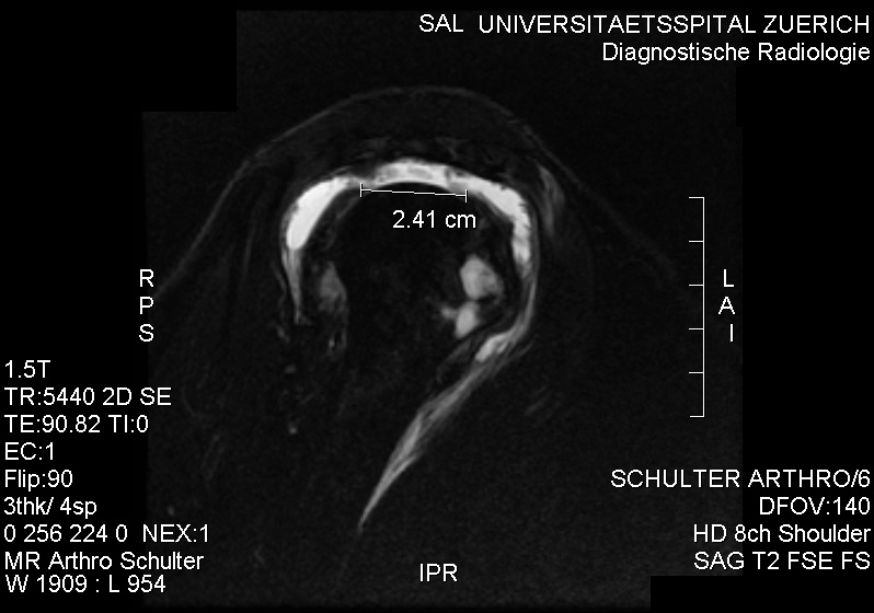
Aims
- Decompress the subacromial space to allow free passage of tendons
- repair tendon back to it’s anatomical position
- ± excision outer clavicle
- Biceps tenodesis
- AC joint
- Chosen over arthroscopic technique for technical ease & better repair.
- Other options. – arthroscopic SAD + mini open repair
Indications
- Full or near full thickness tear usually of supraspinatus tendon
- Significant pain
- Loss of function
- Failed nonoperative management
Contraindications
- Massive tear (>5cm)
- Localised sepsis
Consent / Preop Planning
- Plain radiograph
- U/S or MRI
- Neers test +
- Exclude
- Frozen shoulder
- Cervical radiculopathy
- AC joint Osteoarthritis
- Instability in young patient
- Consent
- Deltoid detachment
- Axillary nerve palsy
- Unable to repair / Repair failure
- Loss of movement / Loss ROM / stiffness
- Persistent pain
- Weakness
- Synovial fistula
Principles
- Antero-inferior acromioplasty
- Release of coracoacromial ligament
- Complete release & mobilisation of cuff from glenoid labrum & superficial bursa ± coracoid base (coracohumeral ligament), rotator interval, & posterior interval (scapular spine)
- Tendon-grasping suture placement (eg. Mason-Allen suture)
- Secure bone fixation (eg. transosseous sutures, suture anchors)
- Minimal deltoid surgical insult & meticulous repair
- Early restoration of passive motion
Options
- Open / miniopen / arthroscopic
- Fixation options:
- transosseous sutures, suture anchors
- absorbable, nonabsorbable
- Single row, double row
Position
- Semi beach chair position, Lateral decubitus
- Arm drape free
Landmarks
- clavicle, AC joint, acromion, spine of scapula, coracoid process
Incision
- Make a 6 cm incision along langers line from the lateral aspect at the middle of the acromion towards & just short of the coracoid
Superficial Dissection
- Undermine skin edges & identify the anterior raphe in the deltoid. Divide this for a distance not exceeding 5cm to avoid the axillary nerve
Deep Dissection
- Carry the dissection across the acromion, subperiosteally elevate off the acromion, ensuring full thickness flaps for repair
Dangers
Nerves
- Axillary nerve
- Musculocutaneous nerve
Procedure
- Resect a 1cm segment of C-A ligament
- Resect thickened bursa (identifiable by unilaminar appearance & attachment to acromion)
- Use rongeur or saw to excise that part of acromion projecting anterior to the anterior border of clavicle
- Place blunt Hohmaan or Bristow to depress humeral head and resect deep part of anterior acromion, half thickness or 7mm at front & tapering back to middle of acromion
- Smooth with rasp
- Identify the tear in tendon. Debride edges
- Gouge a trough at point of insertion
- Non-absorbable sutures through tendon, then through bone
- Check integrity of repair & free passage through sub acromial space
- If deltoid attachment is thin or tenuous, it should be repaired through bone using heavy non absorbable sutures
- Routine closure
Postop / Rehab
- Immediate start of pendula exercises
- 0-6/52 Sling
- 6-10/52 Passive assisted
- after 10/52: as tolerated
Warn patient it may take up to 6 months for pain to subside
Results
- 85-90% pain relief with acromioplasty
- Slight ↑ pain relief with repair of tendon
- Unpredictable ↑ strength with repair
Complications / Dangers
Perioperative
General
- Anaesthetic, general surgical
Local
- Unable to repair tendon
Postoperative
Early
- Infection
- RSD
Late
- Continued pain
- Weakness
Reviewed by Professor Phong Tran, Head of Orthopaedic Surgery, Western Health. Last updated 10 October 2026.