
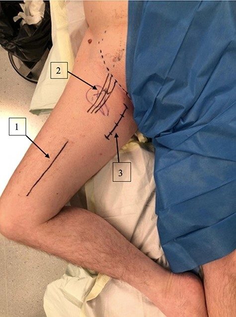
| Position | Supine, hip flexed, abducted and externally rotated |
| Skin | 3-4 cm incision parallel and 1cm distal to the groin crease |
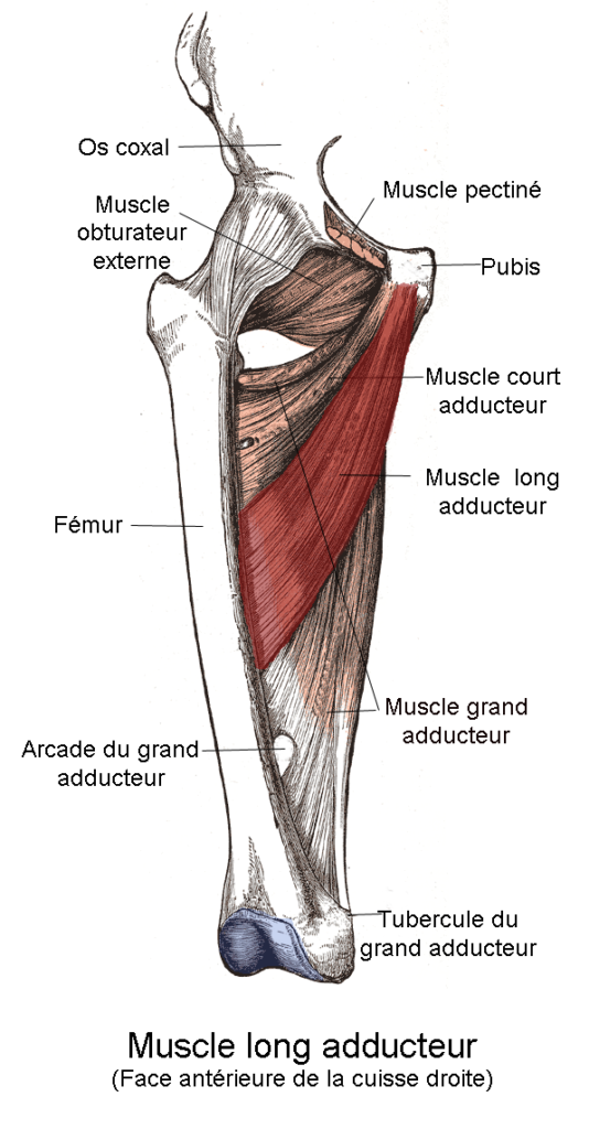
| Superficial | Adductor longus is divided close to the pubic tubercle Obturator nerve (anterior branch) is identified and protected |
| Deep | Between pectineus and adductor brevis Medial Femoral Circumflex Artery is identified and protected Psoas tendon divided at insertion T shaped capsulotomy |
| Dangers | Obturator nerve – predominantly anterior Femoral neurovascular bundle Medial Femoral Circumflex Artery |
| Additional | Divide Transverse acetabular ligament Capsular plication Ligamentum teres tenodesis |
Reviewed by Professor Phong Tran, Head of Orthopaedic Surgery, Western Health. Last updated 10 October 2026.