
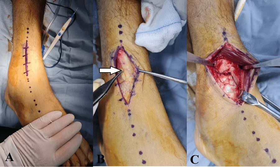
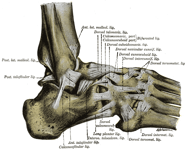
The anterolateral approach to the ankle and hindfoot uses the plane between the extensor tendons and the peroneal tendons to expose the ankle, talonavicular, calcaneocuboid and subtalar joints. It is used for ankle, triple and pantalar arthrodesis, protecting the superficial peroneal nerve.
Aims
- Plane between extensors & peroneal
Indications
- Exposure of
- Ankle joint
- TN joint
- CC joint
- TC joint
- Arthrodesis
- Ankle
- Triple
- Pantalar
- Reduction talus or excision
Position
- supine
- large sandbag under buttock
- tourniquet
- prep & drape
Landmarks
- Lateral malleolus
- Base of 5th metatarsal
Incision
- 15 cm slightly curved longitudinal incision over anterolateral aspect of ankle (2 cm anterior to anterior border of fibula)
- Start: 5 cm proximal to ankle joint
- End: base of 4th metatarsal
Internervous Plane
- Peroneal muscles
- Superficial peroneal nerves
- Extensor muscles
Superficial Dissection
- Incise fascia (no skin flaps)
- Taking care to identify & preserve
- Dorsal cutaneous branches of superficial peroneal nerve
- Taking care to identify & preserve
- Incise superior & inferior extensor retinaculum
- Identify peroneus tertius & EDL
- In upper half of wound
- Incise down to bone just lateral to these muscles
- In upper half of wound
Deep Dissection
- Retract extensor muscles medially to expose the anterior aspect of the distal tibia & ankle joint capsule
- Distally
- Identify EDB at origin from calcaneus
- Detach by sharp dissection
- Cauterise branches of lateral tarsal artery to prevent haematoma formation
- Reflect EDB distally & medially
- Identify the capsules of CC & TN joints
- Identify the fat in the sinus tarsi (laterally)
- Reflect it distally to expose the subtalar joint
Dangers
- Nerves
- Deep peroneal nerve
- Vessels
- Anterior tibial artery
Reviewed by Professor Phong Tran, Head of Orthopaedic Surgery, Western Health. Last updated 10 October 2026.