
Indications
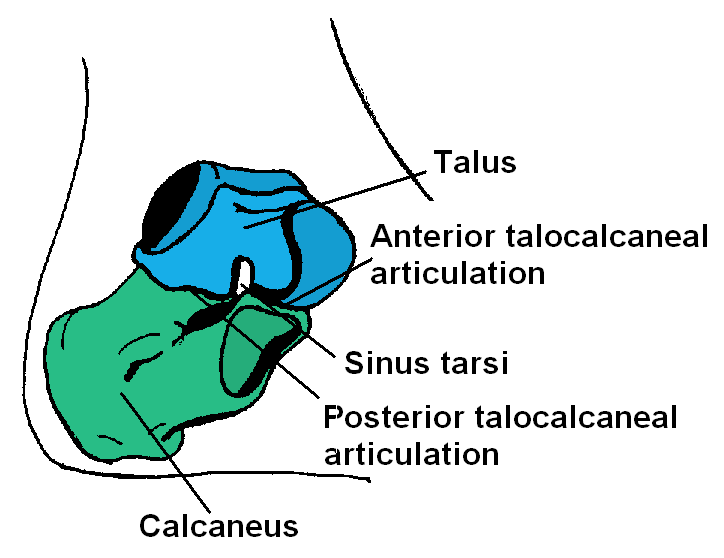
- Subtalar fusions
- ORIF Calcaneal fractures
Position
- supine
- sandbag under buttock to bring lateral malleolus forward
- lateral support on opposite iliac crest
- tilt table 20°
Landmarks
- Lateral malleolus
- Achilles tendon
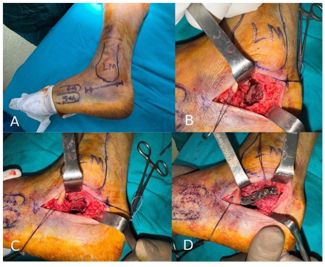
Incision
- 15cm Curved L shaped incision over the lateral aspect of the ankle
- Start
- vertical limb is centred midway between the posterior border of the fibula & anterior border of the achilles tendon
- horizontal limb is centred at the intersection with the thick smooth skin on the sole of the foot & the wrinkly skin on the dorsum of the foot
Internervous Plane
- No internervous plane
- As it reflects both peroneal tendons anteriorly
Superficial Dissection
- Continue my incision straight down to bone achieving thick skin flaps
- Being careful not to damage
- Sural nerve
- Short saphenous vein
- Incise the inferior peroneal retinacula
- Mobilise the peroneal tendons & reflect anteriorly
Deep Dissection
- identify the subtalar joint & overlying calcaneofibular ligament
Dangers
- Skin
- Nerves
- Sural nerve
- Vessels
- nil
Reviewed by Professor Phong Tran, Head of Orthopaedic Surgery, Western Health. Last updated 10 October 2026.