
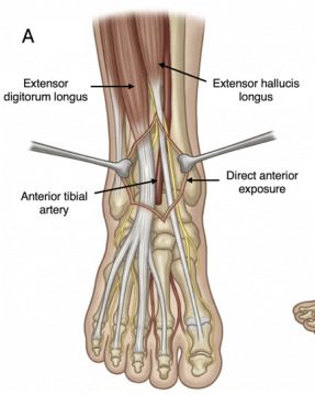
The anterior approach to the ankle joint passes between tibialis anterior and extensor hallucis longus to expose the anterior tibia and talus. It is used for ankle arthrodesis, pilon fracture fixation and joint debridement, protecting the superficial peroneal nerve and the anterior tibial neurovascular bundle.
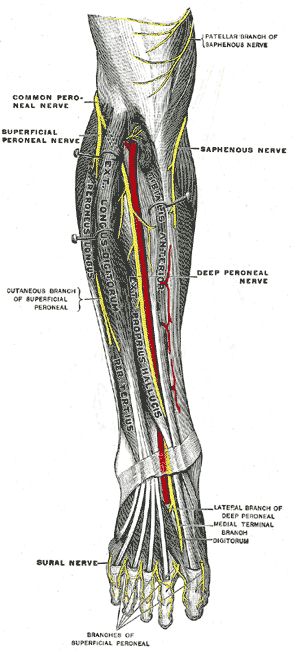
Indications
- arthrodesis
- septic ankle
- r/o loose bodies
- ORIF pilon Fracture
Position
- supine
- exsanguinate foot (partially)
- inflate tourniquet
Landmarks
- medial malleolus
- lateral malleolus
Incision
- Start: 15 cm incision centred over anterior aspect of ankle
- Start 10 cm proximal to joint
- End: dorsum of foot
- Cut only skin
- Bw anterior neurovascular bundle
- Superficial peroneal nerves
Internervous Plane
- None
- Between EHL & ED
- Both supplied by Deep peroneal nerve
- Both receive nerve supply very proximal
Superficial Dissection
- Incise deep fascia
- Incise extensor retinaculum
- Dissect between EHL & ED
- Identify neurovascular bundle just medial to EHL
- Anterior tibial artery
- Deep peroneal nerve
- Retract EHL & NV bundle medially
- Retract ED laterally
Deep Dissection
- Incise ankle joint capsule
- Perform periosteal stripping if necessary
Dangers
- Nerves
- Branches of superficial peroneal nerve
- Deep peroneal nerve
- Artery
- Anterior tibial artery
- NB
- Above ankle joint
- NV bundle lies between EHL & TibAnt
- At joint
- EHL tendon crosses NV bundle
- Above ankle joint
- Proximally
- Expose structures of anterior compartment
- Distally
- Dorsum of Foot
Reviewed by Professor Phong Tran, Head of Orthopaedic Surgery, Western Health. Last updated 10 October 2026.