
Indications
- Most used approach for hip arthroplasty (Moore or Southern)
- Total hip replacement and hemiarthroplasty
- Revision hip arthroplasty
- Open reduction of posterior dislocation
- Drainage of sepsis and removal of loose bodies
- Pedicled muscle grafting and some femoral head procedures
Position
- Lateral decubitus with the affected side up
- Pelvic supports hold the pelvis vertical and stable
- Limb draped free
Landmarks and Incision
- Palpate the greater trochanter and femoral shaft
- Curved incision centred on the posterior trochanter, from the shaft line curving towards the PSIS
- Hip flexion during the incision straightens the curve
Internervous Plane
- No internervous plane
- Gluteus maximus split in line with its fibres
- No denervation, as the inferior gluteal nerve enters well medial to the split
Superficial Dissection
- Fascia lata incised along the femur and continued proximally through gluteus maximus fibres
- Retractors placed under gluteus maximus and fascia lata
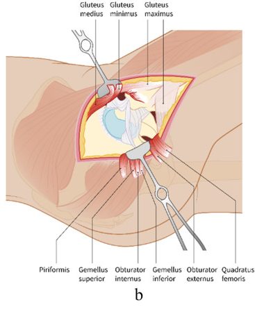
- Trochanteric bursa swept off to expose the short external rotators
Deep Dissection
- Internal rotation tensions the short external rotators
- Piriformis and conjoint tendon detached near their insertions and reflected posteriorly over the sciatic nerve
- Reflected rotators act as a protective layer for the nerve
- Proximal quadratus femoris may be released, controlling the ascending MFCA branch
- Capsule incised, often as a tagged posterior flap
- Dislocation by flexion, adduction and internal rotation
- Repair of capsule and rotators to the trochanter lowers the dislocation rate
Structures at Risk
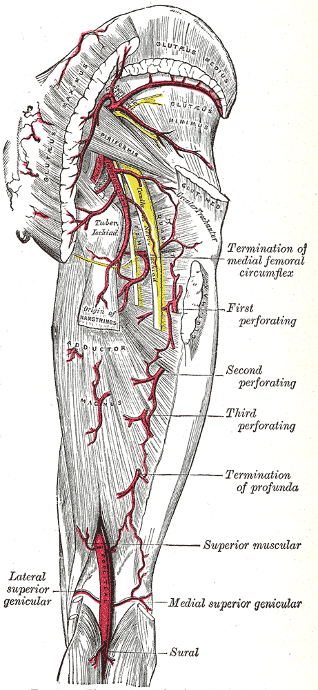
- Sciatic nerve, lying on the short rotators and quadratus femoris
- Inferior gluteal artery below piriformis, which retracts into the pelvis if divided
- Medial femoral circumflex artery near quadratus femoris
- Superior gluteal vessels if dissection strays above piriformis
- Femoral vessels and nerve from aggressive anterior acetabular retractors
Extension
- Distal along the posterolateral femur by elevating vastus lateralis
- Gives whole shaft access for revision, extended trochanteric osteotomy and periprosthetic fracture fixation
- Proximal extension limited by the superior gluteal neurovascular bundle
Reviewed by Professor Phong Tran, Head of Orthopaedic Surgery, Western Health. Last updated 10 October 2026.