
Question
A 36 year old woman has a painless lump on the radial side of the volar wrist that has been present for six months.
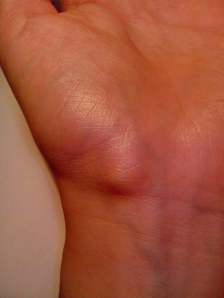
- What is the likely diagnosis and how is it confirmed?
- Where do these lesions arise and what structures are at risk?
- Discuss management and outcomes.
Answer
Diagnosis
- Volar wrist ganglion, second most common site after dorsal
- Smooth, firm lump that transilluminates
- Ultrasound confirms a cyst and shows the relation to the radial artery
- Allen test before any intervention
Origin and structures at risk
- Arises from the radiocarpal or scaphotrapezial joint
- Lies between FCR and the radial artery, often wrapped around the artery
- Radial artery injury during aspiration or excision
- Palmar cutaneous branch of the median nerve
- Superficial radial nerve branches
Management
- Reassurance, about half resolve spontaneously
- Aspiration has a high recurrence rate and risks the radial artery
- Excision for pain or patient choice, include the stalk and a cuff of capsule
- Arthroscopic excision is an option
- Recurrence after open excision of volar ganglia 10 to 30%
- Complications include radial artery injury, scar sensitivity and neuroma
Related pages
Author Contributions
Orthofracs team
Reviewed by Professor Phong Tran, Head of Orthopaedic Surgery, Western Health. Last updated 10 October 2026.