
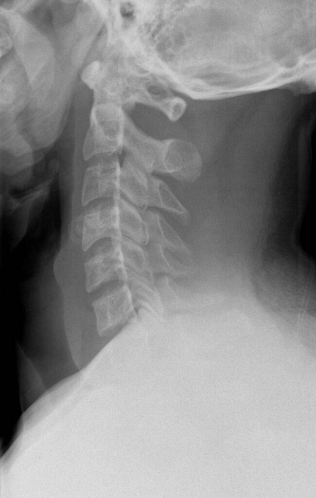
Summary
- Look at posture, alignment and gait
- Feel bony landmarks and paraspinal muscles
- Move through flexion, extension, lateral flexion and rotation
- Upper limb tone, power, reflexes and sensation
- Upper motor neurone signs including Hoffmann sign and clonus
- Special tests including Spurling test and shoulder abduction relief
- Lower limb neurology and gait for myelopathy
Look
- Looking for Asymmetry:
- Fracture malunion, nonunion
- Scoliosis
- Muscular – Torticollis
- Tumours, Glands
Anterior
- Landmarks
- Hyoid C3, Thyroid C4/5, Cricoid C6
- Sternum
- Excavatum, Carinatum
- Muscles
- Sternocleidomastoid
- Torticollis + facial asymmetry
- Sternocleidomastoid
- Syndromes – Marfans, Turners
- Axilla for freckles
- Dysmorphic facies
- Comment on height
- Marfan’s
- skeletal dysplasia (Down’s, achondroplasia, etc.)
Lateral
- Cervical Lordosis
- Thoracic Kyphosis (Normal 21-33)
- in Scheuermann’s, Ankylosing Spondylitis, Fracture
- Gibbus
Posterior
- Skin lesions (hairy naevus, café au lait, neurofibromas)
- Wasting
- Hair line – Klippel-Feil
- Neck webbing
- Shoulder height – Sprengels
- List
Gait
- Ataxic
- e.g. Arnold Chiari or posterior column
- Broad based, halting
- Spastic
- Heel walk, toe walk
- Heel-toe walk for myelopathy/Cb disease
- Rhombergs – posterior column
- -ve with eyes open, +ve with eyes shut
- cerebellar
- +ve with eyes open & closed
Feel
- From Posterior
- Deformity, Muscle Spasm, Tenderness
- Spinous processes (C2 is first palpable)
- Paravertebral muscles
Move
Sitting on Examination Couch
- Mid-range pain suggests instability of moving segment (Degen.)
- Flexion
- chin to chest, measure distance off chest
- Extension
- face parallel to ceiling
- (60% of F/E occurs at Occ/C1)
- Lateral Rotation
- Normal=60-90, 50% occurs at C1/2
- Lateral Bending
- touch ear to shoulder
Neurological Examination
Tone, Clonus
- elbow flexion / extension + pronation / supination
- clasp-knife
Power (Motor)
- Shoulder Abduction (C5)
- Elbow
- Flexion (C6)
- Extension above head (C7)
- Pronation – C7/8
- Supination – C6
- Wrist
- Extension (C6)
- Flexion (C7)
- Fingers
- Extension (C7, C8)
- Flexion (C7, C8)
- Abduction/adduction (T1)
Nerve Roots
- C5 Deltoid
- Comes out between C4/5
- C5/6 Biceps
- C6 Wrist Extensor
- Most commonly affected by disc
- C7 Triceps
- Wrist flexors
- Finger extensors
- C8 Long finger flexors
- Exits between C7/T1 foramen
- T1 Interossei
- i.e. finger abduction/adduction including 1st dorsal
Reflexes
- Biceps – C5, C6
- Triceps – C7
- Brachioradialis – C5, C6
- inverted supinator jerk
Sensation
- C5 lateral arm over the deltoid
- C6 thumb and lateral forearm
- C7 middle finger
- C8 little finger and ulnar border of hand
- T1 medial elbow and upper forearm
- Test light touch and pinprick, comparing sides
- Glove distribution loss suggests myelopathy or peripheral neuropathy
Special Tests
Myelopathy
- Rapid grip/release – 20 times in 10 seconds
- Finger escape test
- Hoffman’s sign – flick terminal phalanx of IF/MF » thumb IP joint flexes
- Do LL’s if myelopathy
- Scapulohumeral reflex for C1-C3 compression
- tap acromion
- +ve if
- arm abduction / scapular elevation
- +ve if
- tap acromion
- Jaw jerk – ↑ in motorneurone disease but not myelopathy
Radiculopathy
- Spurling’s test
- extension + lateral flexion to side of nerve root
- compression & pain
- extension + lateral flexion to side of nerve root
- Shoulder abduction relief test
- decreases radicular pain
Klippel-Feil
- Hands
- polydactyly, syndactyly, thumb hypoplasia, synkinesis
- Scoliosis
- Hair line
- Shoulder height
- C-spine ROM ↓
Reviewed by Professor Phong Tran, Head of Orthopaedic Surgery, Western Health. Last updated 10 October 2026.