
Examination of the foot and ankle includes standing alignment from front and back, gait, heel rise, palpation of bony and tendon landmarks, motion at the ankle, subtalar and midfoot joints, and neurovascular assessment, with special tests for instability and tendon dysfunction.
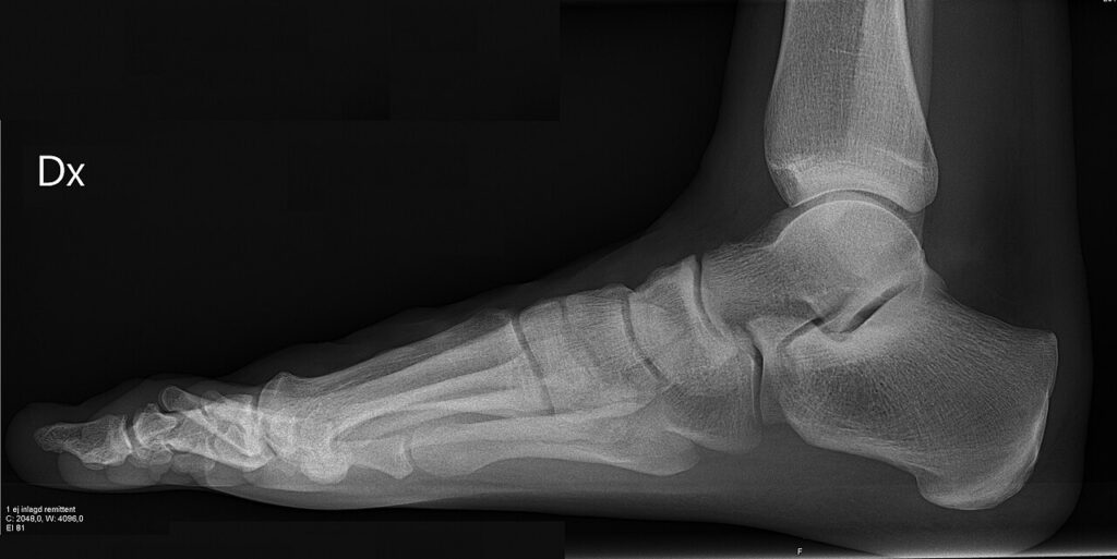
Summary
Ankle and foot consists of 26 bones and 33 joints with many involved muscles, tendons and ligaments.
Look
General Inspection
- Adequate exposure,
- Patient should ideally be wearing shorts to assess ankle, knee and hip as well as a shirt able to assess the back.
- General
- RA, diabetes, evidence of systemic disease
- Mobility aids, external appliances
- Shoes
- Wear pattern
- Off the shelf commercial shoes with no modifications & orthotic
Gait
Ask patient to walk
- Walk away then walk towards
- Normal move of foot through walking – heel strike to toe off
- Gait
- Antalgic (shortened stance phase compared to swing phase)
- Cavus (walking on outside of foot)
- Planus (walking on inside of foot)
- Foot drop (high stepping stance)
- Hallux Rigidus (unable to toe off)
(Gait videos here)
- Foot progression angle
- Angle of foot compared to straight line extending forward from middle of body)
- Slight external rotation normal angle with walking
- Features
- Stiff ankle
- Foot drop
- Fixed equinus
- Antalgic
- Rocker
Wall
Get patient to face wall
Double Heel Raise
- Ability
- Neurological
- likely central cause
- cerebral palsy, spina bifida, cord injury
- Muscular
- muscular dystrophy
- disuse
- Heel movement
- symmetrical heel swing into varus
- heel remains in valgus (abnormal)
- Medial arch
- restoration
- Single heel raise
- on normal foot
- on abnormal foot
- Neurological
- central – stroke, polio, radiculopathy
- peripheral – nerve injury
- Tendinous
- tibialis posterior dysfunction
From Front
Stand patient facing you, describe proximal to distal
Deformity
- Knee alignment: physiological valgus, patella alignment, valgus with pes planus, rotational profile
- Foot rotation: symmetrical ER
- Forefoot: splaying
- Hallux: valgus / varus, pronation, lPJ,
- Lesser toes: overriding, Hammer(PIPJ flexion)/Claw (hyperextension of MTPJ, flexion PIPJ/DIPJ)/Mallet (flexion DIPJ), callosities over IPJ
- Cavovarus deformity of foot exposes heel to anterior view
Skin
- Scars, trophic changes, venous stasis, circulatory disturbance, hair loss, hyperpigmentation, varicosities, ulcers, disuse / denervation, dry skin, dekeratinisation, nails
- Examine between toes
Contour
- Swelling
- Anterolateral ankle
- Navicular
- Bunion
- Bunionette
- Muscle Wasting
- EDB
- Quadriceps
From Medial Side
Turn Affected Side Away
Ensure to assess medial and lateral sides of both feet
Assess Arch
- Pes planus (flat foot)
- Can dorsiflex big toe, if flexible flat foot then arch will reform (Jack test)
- Pes cavus (high arch)
Skin
- See above
Contour
- Swelling
- Tibialis posterior
From Lateral Side
Deformity
- Knee
- Flexion attitude
- Ankle
- Equinus
Skin
- Scars
Contour
- Swelling
- Peroneal tendons
From Behind
Deformity
- Back
- Sagittal spinal deformity
- Cutaneous manifestations of spinal dysraphism
- Lower limb
- rotation/angulation deformity (eg coxa/genu valgum/varum)
- Hindfoot
- Physiological valgus (~5o valgus)
- Varus
- then ask for Coleman Block
- Valgus
- Single heel raise
- Should only be able to 1 and half toes on review posteriorly, any more indicates likely planovalgus
Skin
- See above
Contour
- Swelling
- Calf
- Duchenne
- Tendo Achilles
- Heel
- Haglund’s
- Wasting
- Calf
Sole
Sit patient on side of bed with you seated, inspect sole
Skin
- Scars
- Footprint
- normal
- well-distributed weight-bearing
- heel, lateral border, metatarsal heads
- abnormal
- altered weight distribution
- callosities
- MT heads (II, III), midfoot
- normal
- Lesions
- warts
- ulcers
- soft corns
- pedal sepsis
Contour
- Swelling
- plantar fibromatosis
Feel
Ask Patient “Where Is It Painful?”
Compare temperature of ankle joints
(Add anatomy slides to demonstrate locations of palpation)
Palpation
Sole
- metatarsal heads
- I – sesamoids
- II-V – metatarsalgia
- interdigital neuroma
- interdigital tenderness
- lateral compression – pain & click
- heel
- origin of plantar fascia
Medial
- 1st Metatarsal head
- 1st metatarsal-Cuneiform joint (flare of base of 1st MT)
- Navicular Tubercle
- Head of Talus (evert forefoot)
- Medial Malleolus
- Sustentaculum Tali (1.5 cm below medial malleolus)
- Posterior impingement/ Os trigonum
- palpate posterior to medial malleolus and dorsiflex to reproduce pain
Lateral
- 5th Metatarsal head
- Cuboid
- Calcaneum
- Tender in this region in Haglund’s deformity (overgrowth of bone around Achilles insertion)
- Peroneal Tubercle (separates peroneus longus & brevis)
- Lateral malleolus
Tendons
- Tendo Achilles
- insertion
- tenderness
- along tendon
- tenderness (diffuse or focal)
- lump
- retrocalcaneal bursa (pinch tissue anterior to AT)
- calcaneal bursa (posterior to AT)
- insertion
- Tibialis Posterior
- prominent with plantarflexion & inversion
- thickening
- tenderness
- crepitus
- check power if abnormal
- navicular insertion
- Peroneals – Brevis & Longus
- thickening
- tenderness
- dislocation (resisted eversion)
- base of 5th MT insertion
- Tibialis Anterior
- Prominent with dorsiflexion & inversion
- Insertion
- EHL & EDL
- Dorsiflex toes
Ankle Joint
- Lateral ligaments
- 1. Anterior Talo-Fibular Ligament (ATFL)
- 2. Calcaneo-Fibular Ligament (CFL)
- 3. Posterior Talo-Fibular Ligament (PTFL)
- 4. Anterior Inferior Tibiofibular Ligament (AITFL)
- 5. Posterior Inferior Tibiofibular Ligament (PITFL)
- Joint line
- Deltoid Ligament (Medial ligament)
- 1. Anterior Tibio-Talar Ligament
- 2. Tibio-Calcaneal Ligament
- 3. Posterior Tibio-Talar Ligament
- 4. Tibio-Navicular Ligament
- Deep
- Superficial
- Fluctuance
- Effusion or synovial thickening
Forefoot
- Bunion
- Bunionette
- Pulses
- Dorsalis pedis
- Posterior tibial
- Capillary refill
- Neurological
- SPN- sensation: across forefoot, motor eversion of foot
- DPN- sensation: 1st webspace, motor extension of hallux/phalanges
- TN- sensation: sole of foot (medial and lateral sides different branches of tibial nerve); motor: flexion of hallux/phalanges
- Sural- sensation along lateral aspect of foot
(Insert picture of pulses/neurology/lower limb compartments)
Move
- Active, Passive, Power
- Compare both feet at same time
- Dorsiflexion
- Plantarflexion
- Inversion
- Eversion
- Ankle / Tibiotalar (Dorsiflexion/Plantarflexion)
- thumb on talar neck
- grasp heel with other hand
- plantarflex & dorsiflex ankle
- movement occurring at ankle joint
- relate to foot at neutral (90°)
- dorsiflexion 20°
- plantarflexion 50°
- Subtalar (Inversion/Eversion)
- forefoot held in neutral at 90° to tibia (locks wider, anterior part of talar dome into ankle mortise)
- foot relaxed with thumb on talar neck
- grasp heel with other hand
- invert & evert heel
- inversion 10-15°
- eversion 0-5°
- Tarsal Coalition
- Restricted subtalar motion
- Midtarsal
- (Adduction/Abduction; Dorsiflexion/Plantarflexion)
- Foot held at 90° (to lock talus into ankle mortise)
- Calcaneus grasped
- Adduct foot (20°)
- Abduct foot (10°)
- Dorsiflexion
- Plantarflexion
- 1st MTPJ (Flexion/Extension)
- Stabilise forefoot & IPJ
- extension 70-90°
- flexion 45°
- 1st IPJ (Flexion/Extension)
- stabilise proximal phalanx
- flexion 90°
- extension 0°
- Toes (Flexion/Extension)
- extension only occurs at MTPJ
Special Tests
Simmond’s Test
- With patient kneeling/lying prone
- Squeeze calf
- Plantar flexion should occur
- If nil movement, suspect Tendo Achilles Rupture
Instability
- Anterior Drawer
- Due to complete tear of ATFL
- grasp lower tibia & cup calcaneum
- “clunk” or draw
- cf. other side
- Lateral instability
- Inversion stress
- gaping of soft tissues
- talar tilt (may occur in normal & must cf. with other side)
- needs to be confirmed on stress views
- Medial Instability
- Eversion stress
- gaping/ widening
- needs to be confirmed on stress views
Gastrocnemius / Soleus Contracture
- test if limited dorsiflexion
- extend knee – dorsiflexion limited by both soleus & gastrocnemius contracture
- flex knee – gastrocnemius relaxed (crosses knee joint)
- if dorsiflexion still limited it is due to soleus contracture
- if limited in extension & not in flexion then due to gastrocnemius contraction
Pes Cavus
- Modify
- claw toes
- individual power cf. other side
- tibialis anterior (inversion in DF)
- tibialis posterior (inversion in PF)
- peronei
- Add
- Coleman block test
- dynamic visualisation of hindfoot correction
- stand on 2cm block
- Sensation
- Spine
- Hands
Hallux Valgus / Rigidus
- Add
- MTPJ
- dorsal osteophytes
- passive ROM
- attempt to correct deformity
- grind test
- hallux interphalangeus
- lesser toes
Lesser Toes
- Add
- describe deformity
- claw, hammer, mallet, overriding, curly
- callosities
- palpate joints
- MTP, PIP, DIP
- fixed or mobile
- subluxed or dislocated
Adult Flatfoot
- Modify
- tibialis posterior function
- Lisfranc joint
Tarsal Coalition
- Modify
- palpate
- medially (sustentaculum tali)
- dorsolaterally (through EDB)
Lateral Ligament Instability
Add
- Anterior drawer
- Talar tilt
Reviewed by Professor Phong Tran, Head of Orthopaedic Surgery, Western Health. Last updated 10 October 2026.