
Definition
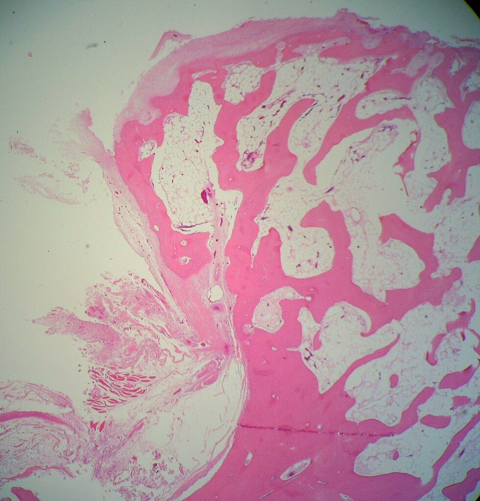
- Cartilage capped bony projection
- Cortex and medulla continuous with the host bone
- Commonest benign bone tumour
- Solitary, or multiple in hereditary multiple exostoses (HME)
Aetiology
- Displaced physeal cartilage growing at an angle to the bone
- Grows by enchondral ossification, stopping at skeletal maturity
- HME is autosomal dominant, from EXT1 or EXT2 mutations affecting heparan sulphate synthesis
- EXT1 mutations cause more severe disease
- Radiation induced lesions follow childhood radiotherapy
Clinical Features
- Painless hard lump near a joint in childhood or adolescence
- Distal femur, proximal tibia and proximal humerus metaphyses
- Points away from the adjacent joint
- Pedunculated or sessile
- Mechanical irritation with an overlying bursa
- Common peroneal nerve compression at the fibular neck
- Popliteal artery pseudoaneurysm
- Fracture through the stalk or restricted joint movement
- HME causes short stature, ulnar shortening, radial bowing and radial head dislocation
- HME also causes genu valgum and ankle valgus
Investigations
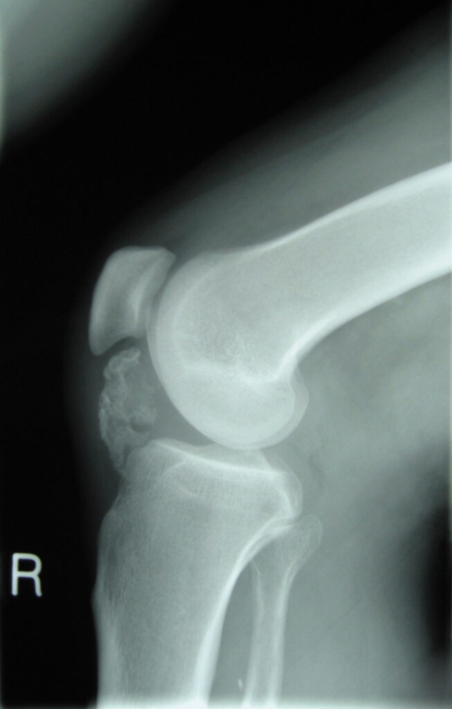
- Radiographs show corticomedullary continuity and are usually diagnostic
- MRI or CT for atypical, pelvic or scapular lesions and cap thickness
- Ultrasound measures the cap in superficial lesions
Complications
- Secondary peripheral chondrosarcoma, usually low grade
- Risk around 1% or less for solitary lesions, a few percent lifetime in HME
- Pelvis, shoulder girdle and proximal femur carry greater risk
- Warning signs are growth after maturity and new non-mechanical pain
- Cap thickness over 2 cm in an adult
- Irregular cap margin, scattered calcification or soft tissue mass
Management
- Observe asymptomatic lesions
- Excise at the base with the entire cap and perichondrium
- Delay excision until near skeletal maturity to reduce recurrence
- Protect the physis and neurovascular structures
- HME may need guided growth, ulnar lengthening, osteotomy or radial head excision
- Suspected chondrosarcoma goes to a sarcoma centre for wide resection, often without prior biopsy
- Adults with HME report new pain or growth
Reviewed by Professor Phong Tran, Head of Orthopaedic Surgery, Western Health. Last updated 10 October 2026.