
Anatomy
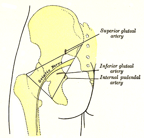
- L4 to S3, exits greater sciatic foramen usually below piriformis
- Peroneal division may pass through or above piriformis
- Lies on adductor magnus deep to the long head of biceps
- Divides near the popliteal fossa apex, though at any level
- Peroneal division is lateral and more vulnerable
- Fewer larger fascicles, less connective tissue, tethered at notch and fibular neck
- Short head of biceps is the only thigh muscle from the peroneal division
Aetiology
- Posterior hip dislocation, mostly peroneal division
- Posterior wall or column acetabular fractures and Kocher-Langenbeck fixation
- THR risk rises with dysplasia, revision, large lengthening and posterior approach
- Haematoma, cement extrusion and retractors in THR
- Injection, gunshot and stab wounds
- Prolonged compression, hamstring avulsion repair, piriformis syndrome and tumours
Clinical Features
- Foot drop is the commonest presentation
- Hamstring weakness indicates a lesion above the knee
- Preserved hip abduction separates it from L5 radiculopathy
- Tibialis posterior weakness and short head of biceps denervation separate it from fibular neck palsy
Investigations
- NCS and EMG at 3 to 4 weeks, including short head of biceps
- Ultrasound or MRI for haematoma or tumour
- CT for screws, cement or fragments
Management
- Reduce hip dislocation urgently
- Hip extended and knee flexed during acetabular fixation
- Post THR palsy prompts review of leg length and component position
- Urgent exploration for expanding haematoma, implant impingement or excessive lengthening
- Otherwise AFO, physiotherapy and neuropathic analgesia
- Explore if no recovery at about 3 months
- Tibialis posterior transfer for persistent foot drop
Prognosis
- Partial lesions and the tibial division recover better
- Recovery may continue up to 2 years
- Complete peroneal lesions often leave permanent foot drop
- Grafting long gaps gives poor results
Reviewed by Professor Phong Tran, Head of Orthopaedic Surgery, Western Health. Last updated 10 October 2026.