
Anatomy
- Endoneurium surrounds each axon
- Perineurium groups axons into fascicles and provides tensile strength and the blood nerve barrier
- Epineurium encloses the fascicles
- Myelinated axons conduct by saltatory conduction between nodes of Ranvier
Classification
Seddon
| Type | Pathology | Recovery |
|---|---|---|
| Neurapraxia | Local conduction block from segmental demyelination, axon intact, no Wallerian degeneration | Full |
| Axonotmesis | Axon and myelin disrupted with the connective tissue framework preserved, Wallerian degeneration follows | By regeneration |
| Neurotmesis | Complete disruption | None without surgery |
Sunderland
| Grade | Injury | Recovery |
|---|---|---|
| I | Neurapraxia, conduction block | Full |
| II | Axon disrupted, endoneurium intact | Full, at the rate of regeneration |
| III | Endoneurium disrupted, perineurium intact | Incomplete, from axonal misdirection |
| IV | Only epineurium intact | Neuroma in continuity needing resection and repair or grafting |
| V | Complete transection | None without repair |
| VI (Mackinnon) | Mixed injury with different grades in different fascicles | Variable |
Original publication Sunderland S. A classification of peripheral nerve injuries producing loss of function. Brain. 1951;74(4):491-516.
Pathology
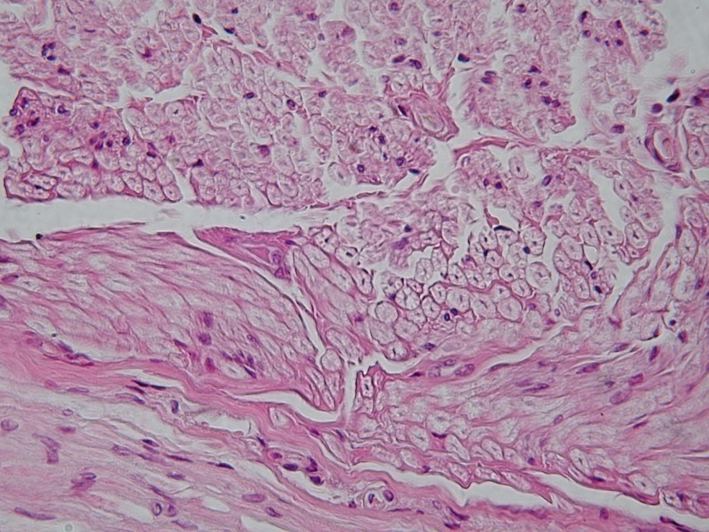
- Distal axon and myelin break down over the first days (Wallerian degeneration)
- Schwann cells align in endoneurial tubes as bands of Büngner, guiding sprouts and secreting neurotrophic factors
- Distal segment conducts for several days after transection, affecting nerve study timing
- Cell body undergoes chromatolysis, swells and increases protein synthesis
- Severe proximal injury can cause neuronal death
- Regeneration runs at about 1 mm per day (1 inch per month), faster proximally
Clinical Features
Tinel sign
- Percussion from distal to proximal produces paraesthesia at the level of regenerating axon tips
- Distal advance on serial examination indicates regeneration
- A static sign at the injury site, stronger than any distal response, suggests neuroma or failed regeneration
- Shows some axons are regenerating but does not quantify them or predict function
Prognosis
- Motor end plates and denervated muscle fibrose irreversibly after about 12 to 18 months
- Sensory receptors tolerate denervation longer, so sensory recovery can occur late
- Better outcome with young age, distal injury and a sharp mechanism over crush or avulsion
- Early, tension-free repair with a short gap does better
Reviewed by Professor Phong Tran, Head of Orthopaedic Surgery, Western Health. Last updated 10 October 2026.