
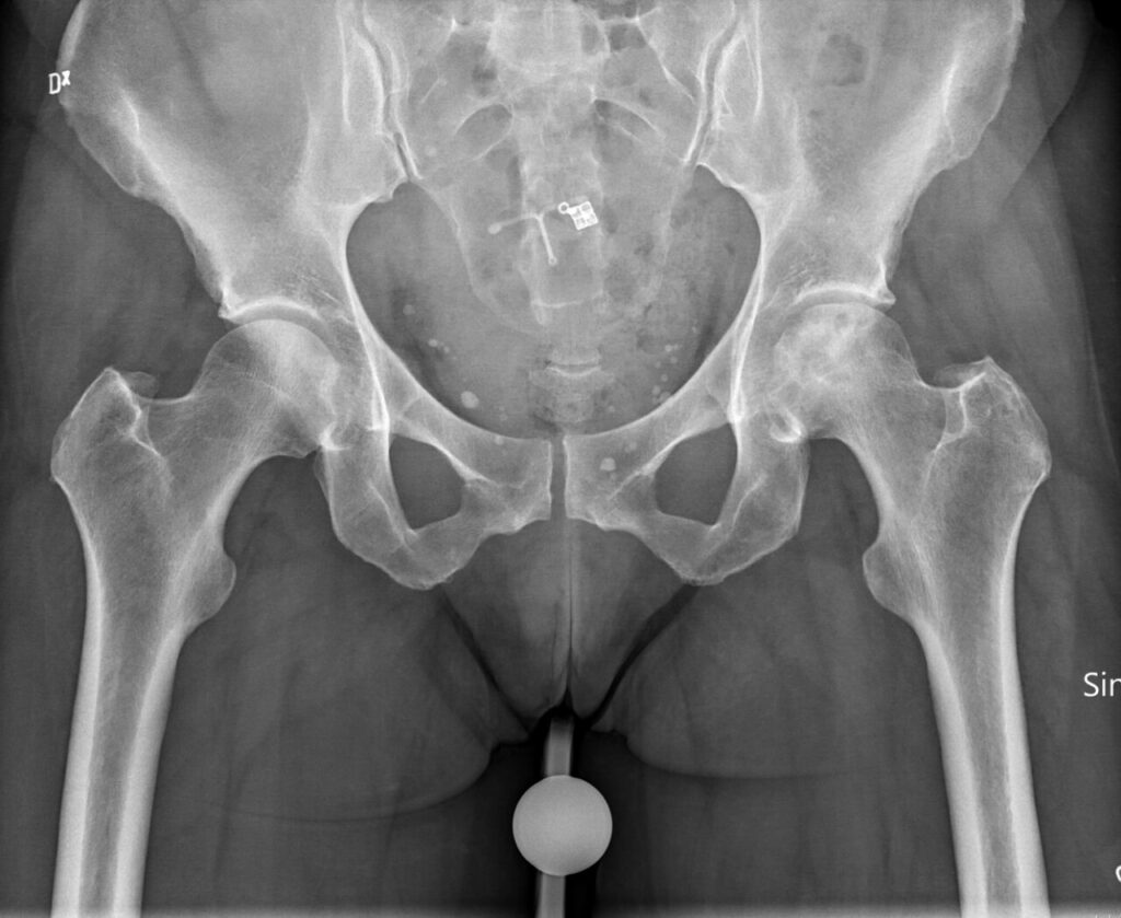
Osteonecrosis (AVN)
Definition
- In situ death of a segment of bone due to ischaemia
Locations
- Femoral Head
- Femoral Condyles
- Humeral Head
- Capitellum
- Lunate
- Scaphoid
- Talus
- ie. Convex bones or small cuboidal bones
- Following surgery
- eg. Head of first metatarsal
Aetiology
- Trauma
- Fractures
- Dislocations
- Non-Traumatic
- Alcohol abuse
- Corticosteroid usage
- Caissons Disease
- Sickle cell disease
- Thalassaemia
- Gauchers disease
- Infection
- Congenital
- medial (multiple epiphyseal dysplasia)
- Developmental
- Perthes disease
- SUFCE
- Idiopathic (most common)
Pathogenesis
- Due to ischaemia of bone
- Numerous theories
- Four mechanisms that are mutual rather than exclusive
- Interruption arterial supply
- Capillary occlusion
- Intraosseous capillary tamponade
- Injury to vessel wall
- 1. Arterial insufficiency
- Fractures & dislocations most often seen
- SUFCE & Perthes related
- NOF
- poor associated collateral blood supply
- DDH
- following treatment
- 2. Intravascular Capillary Occlusion
- Due to vascular sludging
- Caissons Disease
- nitrogen bubbles
- Corticosteroids
- fat emboli
- Alcohol
- fat emboli
- Sickle Cell disease » abnormal RBC
- 3. Intraosseous Capillary Tamponade (intraosseous HTN)
- Osteonecrosis as a compartment syndrome
- Commonest cause of AVN
- Corticosteroids
- enlarged marrow fat cells
- Alcohol
- enlarged marrow fat cells
- Post-infection
- inflammatory response
- also related to activation of intravascular DIC
- Gauchers Disease
- glycocerebroside in bloated macrophages
- 4. Vessel Wall Damage
- DXRT
- radiation-induced vessel disease
- SLE
- vasculitis
- DXRT
- Can also add…
- Venous Occlusion (Chandler’s disease)
- Venular pressure > Arteriolar pressure
- Must be very extensive to produce vascular stasis
- Capsular tamponade from effusion, trauma etc
- Perthes disease
- Infection
Pathogenesis
Stages of the disease process
| Stage | Process | Features |
|---|---|---|
| 1 | Death | Ischaemic event with segmental bone death and opaque yellow marrow. Marrow cells die in 6 to 12 hours and osteocytes in 24 to 48 hours |
| 2 | Inflammation and revascularisation | Capillaries and mesenchymal cells advance from adjacent live marrow into the dead marrow spaces, forming a cuff of vascular granulation tissue |
| 3 | Repair | Macrophages remove dead fat and debris, osteoclasts resorb dead bone and osteoblasts lay new bone on dead trabeculae (creeping substitution), seen as sclerosis. Incomplete healing leaves fibrous tissue and cysts, and collapse can follow stress fracture of dead bone or weakness at the repair front |
| 4 | Remodelling | Dead trabeculae removed and woven bone converted to lamellar bone |
| 5 | Osteoarthritis | Secondary to subchondral collapse and altered force transmission |
Traumatic AVN
- Overlying superficial cartilage to tideline that receives nutrition from joint fluid not involved
- Cartilage below tideline dies due to disruption of blood supply
- The bone below this area also necrotic
- Influx of inflammatory cells & macrophages to remove infarcted marrow
- Then see ingrowth of fibrovascular tissue that differentiates into osteoblasts & deposits new bone on dead trabeculae
- Then replacement of the central necrotic bone – Creeping substitution
- If fibrovascular tissue fails to reach infarct then necrotic marrow undergoes ectopic calcification – MUCH more common in non-traumatic infarcts
Atraumatic AVN
- The differences are related to the fact that the original fibrovascular tissue from first infarct prevents further new ingrowth of mesenchymal tissue after this & so it becomes calcified with repeated infarction
- The area of subchondral bone beneath viable cartilage resorbed & so stress fracture common in this area
- Interior of infarct remains unrepaired
- Subchondral/ Juxta-articular AVN
- Most commonly affects
- Anterosuperolateral portion of femoral head
- Central dome of humeral head
- Infarction of medullary bone & cortex
- Loss of medullary & cortical architecture
- Painful & progressive
- If subchondral collapse
- Crescent Sign
- Secondary Osteoarthritis
- Wrinkle appears at dead margin
- Then fissures appear
- Escape of bony detritus through crack into joint leads to synovitis
- Cartilage may lift off
- Fibrocartilage may form on sequestrum
- Stage 1
- Joint unaltered & external examination of joint shows no abnormalities
- Cut section of necrotic zone shows wedge shaped region with dull-yellow & chalky marrow in subarticular area
- Surrounding marrow separated by thin red hyperaemic border
- Microscopically the articular cartilage is viable down to calcified zone (tidemark)
- The subchondral bone has replaced marrow elements with eosinophilic granular material containing ghosts of fat cells
- Extensive calcification may be present due to repeated AVN episodes
- At margin of infarct is proliferation of osteoblasts & fibroblasts/ capillaries moving into medullary space
- Radiologically not detectable
- Stage 2
- The overall shape of bone intact & articular surface radiologically intact
- However see Sclerotic Rim at the boundary between necrotic zone & unaffected marrow
- Central region of necrosis unchanged but the hyperaemic zone thicker
- Microscopically advancing front of granulation tissue, lipid laden macrophages, fibroblasts & capillaries at periphery & extending into necrotic zone
- A second front at a distance has dead bone being resorbed by osteoclasts – Creeping Substitution (Phemister) » removal of necrotic tissue whilst maintaining structural integrity
- Accounts for ↑ uptake on radionuclide scanning
- Stage 3
- Alteration in bone shape becomes radiologically identifiable
- Collapse in necrotic area occurs
- On gross inspection the trabecular bone is fractured below the bony end plate
- Subchondral fracture follows – Crescent Sign » the cartilage above springs back & lucent line produced
- Trabecular fracture due to
- – Cumulation of fatigue-induced microfractures
- – Weakness of trabecular bone in reparative front due to osteoclastic
- activity
- – Stress risers at the junction of necrotic bone & the reparative front
- Microscopically appears as bony & cartilaginous debris
- Overlying cartilage may still look viable
- Appearance of unstable non-united fractures elsewhere
- Deep trabecular fracture may produce no overlying changes although can see articular depression
- Stage 4
- Morphological changes of degenerative arthritis
- Medullary Osteonecrosis
- Infarction of medullary bone
- Usually caused by medical conditions
- Dysbarism
- Haemoglobinopathies
- Gauchers disease
- Most commonly affects
- Lower femur
- Upper tibia
- Upper humerus
- Variable extent
- Asymptomatic usually
- Silent & non-progressive
- Similar pathology
- Collagen Calcium
- “Coil of Smoke” sign
- Pathology
- Dead marrow yellow & opaque
- Surrounded by dense collagen layer that may be calcified
- Cortical width ↑ if close
Radiology
- Main change is calcification
- Wavy line of ↑ density – Coil of Smoke Sign
- Endosteal cortex thickened
- Difficult to distinguish from
- Bone island
- Calcified enchondroma
- Investigations
- Blood Tests
- Resistance to activated protein C
- Lipoprotein Lp(a)
- Protein C & S
- Tissue plasminogen activator & inhibitor
- Antiphospholipid antibodies
- Plain radiographs
- Mottling
- Sclerotic line at junction of dead bone
- Later
- Crescent sign with joint collapse (best seen in frog leg lateral of femoral head)
- Segmental collapse
- End-stage changes of Osteoarthritis
- Until Osteoarthritis joint space is maintained
- Dead bone appears dense due to
- Compression of dead trabeculae
- Subchondral fracture
- Calcium of dead marrow (saponification)
- Onlay of new bone on dead trabeculae
- Relative osteosclerosis with surrounding osteopenia
- Bone Scan
- Initially ↓ uptake
- Doesn’t absolutely predict AVN as revascularisation may occur without necrosis
- Later ↑ uptake due to repair
- Hot later doesn’t necessarily predict good outcome as revascularisation may be inadequate
- Non-specific
- Cold areas may be metastases
- Hot areas may have numerous causes
- Most useful to detect avascularity
- After acute femoral neck fracture or hip dislocation
- CT Scan
- Not useful in early stages
- Best to differentiate precollapse stage 2 from structural collapse of stage 3
- Good to detect
- Extent of subchondral fracture
- Flattening & collapse of articular surface
- MRI
- Gold standard in early detection
- Most sensitive & specific
- Femoral head most extensively studied
- Normal marrow rich in fat » High signal intensity on T1
- Dead marrow » Decrease in signal intensity on T1
- T1 see low signal line
- Earliest
- Avascular-Vascular bone interface
- T2 see double-line
- Outer low signal line is thickened trabeculae
- Inner high signal line is granulation tissue
- Advantages are
- Early detection » pre-radiological
- Accurate localisation & extent of area involved
- Change in signal early related to ↑ water content
- Functional Exploration
- 3-phase invasive investigations – Ficat
- Intraosseous pressure
- Abnormal > 30mmHg
- Intramedullary Venogram
- Biopsy
- No longer used
- Non-Traumatic Osteonecrosis of Femoral Head
- Epidemiology
- Most common in middle aged men
- M:F is 4:1
- Peak incidence is 30-60 years
- Bilateral in 50% of idiopathic & 80% of the corticosteroid-related
- Aetiology
- Exposure to alcohol & steroids make up 90% of cases where aetiology established
- Alcohol
- Most common presentation
- 15-75% of patients
- 5-30% of alcoholics develop AVN
- 50% bilateral
- Studies suggest as little as 400ml/ week is enough
- Most idiopathics likely to be alcohol-related
- Steroids
- Risk related to the length of treatment & size of dose
- Overall risk is 3-25%
- Interval from use to time of onset varies from 6 months to 3 years
- Often multiple sites involved
- Often bilateral (80%)
- Usually progresses to joint failure
- Conditions
- Post-Transplantation
- 20% initially
- Now 2% due to use of Cyclosporin instead
- Usually onset within 1 year but can be up to 6 years
- Involves the
- Femoral head
- Humeral head
- Femoral condyle
- In decreasing order of frequency
- Leukaemia/ Lymphoma
- Rheumatoid
- Asthma
- Differences from traumatic AVN are
- Anterolateral position of lesion in femoral head
- Repetitive nature of lesions compared with single event in traumatic form
- Clinical Features
- Aching pain in groin & thigh
- Radiates to knee & buttocks
- Gradual onset
- Occasionally sudden
- Initially mechanical
- ROM reduced due to pain particularly Internal rotation & Abduction
- Click
Ficat and Arlet staging of femoral head osteonecrosis, with Hungerford and Lennox stage 0
| Stage | Clinical | Imaging |
|---|---|---|
| 0 | Preclinical | Radiographs normal, bone scan cold, MRI double line on T2 |
| I | Onset of ischaemia | Radiographs normal, changes on MRI or bone scan only |
| II | Pain | Early cystic and sclerotic change with a preserved head contour |
| III | Structural change | Crescent sign and flattening of the head |
| IV | Degenerative change | Joint space narrowing and secondary osteoarthritis |
Original publication Ficat RP. Idiopathic bone necrosis of the femoral head. Early diagnosis and treatment. J Bone Joint Surg Br. 1985;67(1):3-9.
- Steinberg extends staging to stages 0 to VI and divides stage III by the presence of collapse
- Steinberg also grades the extent of subchondral fracture as A mild (under 15%), B moderate (15 to 30%) or C severe (over 30%)
- The Japanese Investigation Committee grades location as medial (A), central (B) or lateral (C)
- Enneking (Florida) classification is also described
- Treatment
- NON-Operative
- Observation & Protected Weight-Bearing
- Stage 1 & 2 left untreated will collapse in 85% at 2 years
- Meta-analysis of 21 studies & 819 hips at 3 years
- – 74% had radiological progression
- – 76% required arthroplasty
- – The incidence of radiological progression was related to stage
- time to failure was not related to degree of NWB
- The very small lesions < 15% may heal & not progress with non op treatment
- OPERATIVE
- Core Decompression (Forage)
- Ficat & Arlet 1964
- Rationale
- – Reduction of Intramedullary pressure
- – Stimulate angiogenic & osteoblastic responses (enhances creeping substitution)
- – Pain relief
- – Tissue for diagnosis
- Procedure
- – Fracture table & II
- – Lateral trochanteric approach
- – Hollow biopsy trephine – 8-10mm
- – The anterolateral part of head within 5mm of the articular surface
- – 3mm drill used to penetrate the necrotic segment to subchondral bone
- Results
- Divergence of opinion
- Meta-analysis of 24 studies involving 1206 hips at 3 years
- 37% did not progress radiologically
- 33% required arthroplasty
- Success related to stage
- – Stage 1 – 84%
- – Stage 2 – 65%
- – Stage 3 – 47%
- Two studies compared core decompression with non-op treatment
- No collapse in 61% vs 39%
- No arthroplasty in 75% vs 29%
- Complications
- Uncommon
- Include
- – Subtrochanteric fracture
- – Infection
- Indications
- Stage 1 & 2 disease
- Stage 3 where not suitable for more extensive procedure
- Non Vascularised Bone Grafting
- Cortical bone graft into defect produced with core decompression
- Rationale
- Provides mechanical support for articular surface during healing
- Procedure
- Cortical strut graft from the
- – Ilium
- – Fibula
- – Tibia
- Inserted into core track
- Protected weight bearing for 3-6 months until radiographic evidence of healing
- Results
- Conflicting reports
- Success rates of 60-80% with short term follow up
- Some long term reports have 30% successful outcome
- Disadvantages
- Prolonged restricted weight bearing
- Indications
- Early stage 3 lesions?
- Unsuccessful core decompression
- Vascularised Bone Grafting (Urbaniak 1987)
- Rationale
- To enhance revascularisation so that progression of necrosis altered
- Vascularised grafts undergo more rapid & complete incorporation
- Procedure
- Considerable variability
- Donor site
- – Ilium
- – Fibula
- – GT
- Muscle pedicle artery & vein used
- – Inferior gluteal
- – Profunda femoris
- – Circumflex
- Results
- Most studies have short term follow up in small numbers
- Most comprehensive is Yoo – 81 hips at 5 years
- Vascularised fibula to profunda femoris
- 91% of Stage 2 & 3 had Good-Excellent results
- 89% had no radiological progression
- However the rate of conversion to THR is identical to core decompression at 20%
- Indications
- Stage 2 or early Stage 3 lesion
- Young patient
- Large lesion
- Osteotomy
- Rationale
- Transfer load from necrotic area to undamaged part
- Transection of bone may afford decompression
- Procedure
- Maybe flexion/ extension/ valgus/ varus or rotational
- If superolateral then need
- Flexion
- Valgus
- If central then
- Varus
- Flexion
- Sugioka
- Transtrochanteric rotational osteotomy
- Technically demanding
- Can rotate through 90°
- Poor results if not intertrochanteric with damage to blood supply of the head
- Results
- Sugioka osteotomy in 52 hips Stage 3
- 56-69% at average of 5 years successful
- If > 50% involved then results poor
- Deterioration with time – only 40% of hips surviving 7-10 years
- Cumulative necrotic sector angle (Kerboul)
- Angle from centre of femoral head to edges of necrotic sector measured on AP & lateral films & added together – if < 200° then a favourable outcome after femoral osteotomy may be expected
- Disadvantages
- Make subsequent THR difficult
- Indications
- Stage 3 disease
- Small lesion
- No ongoing cause for AVN
- Hemiarthroplasty
- Poor results – 50% revision rate
- Loosening & Protrusio biggest problem
- Study showed almost universal acetabular cartilage disease at time of arthroplasty
- Total Hip Replacement
- Better results – preferred treatment?
- Failure rate higher than for other diagnoses
- Related to poorer bone stock
- 30-50% revision rates at 10 years
- In < 50 yo with AVN cemented THR (metal on poly) has 50% failure rates at 10 years (Dorr) – apparently no other study gives better results than this
- Arthrodesis
- Usually contraindicated
- As the disease is bilateral in 50-80%
- Reasonable option in young, active, heavy man with unilateral disease
- Electrical Stimulation
- Not proven technique
- May be adjunct to other surgery
- AVN of Humeral Head
- 2nd most common site
- Smoking ↑ risk 4x
- Primary vascular supply
- Anterior circumflex humeral artery
- Arcuate artery once in bone
- 26-75% rate AVN after 4-part fracture
- Avoid activities above shoulder height
- » Greatest joint reaction force
- Benefit of core decompression ambiguous
- 94% relief of pain with prosthesis
Reviewed by Professor Phong Tran, Head of Orthopaedic Surgery, Western Health. Last updated 10 October 2026.