
Definition
- Osteochondritis dissecans is an acquired lesion of subchondral bone with secondary involvement of overlying cartilage
- Risk of fragment instability, separation and loose body formation
- Juvenile form occurs with open physes, adult form after physeal closure
Aetiology
- Uncertain
- Repetitive microtrauma is the leading theory
- Impingement of the tibial spine on the medial femoral condyle proposed
- Ischaemia of subchondral bone, abnormal ossification and genetic factors also proposed
Epidemiology
- Peak in adolescence, males more than females
- Frequently in active children
- Bilateral in a minority, so image the other knee
Anatomy
- Most common on the lateral aspect of the medial femoral condyle
- Less often lateral femoral condyle, trochlea and patella
Clinical Features
- Vague activity related knee pain and effusion
- Mechanical locking or catching suggests an unstable fragment or loose body
- Wilson test, pain on internal tibial rotation during extension relieved by external rotation
- Wilson test has low sensitivity
Investigations
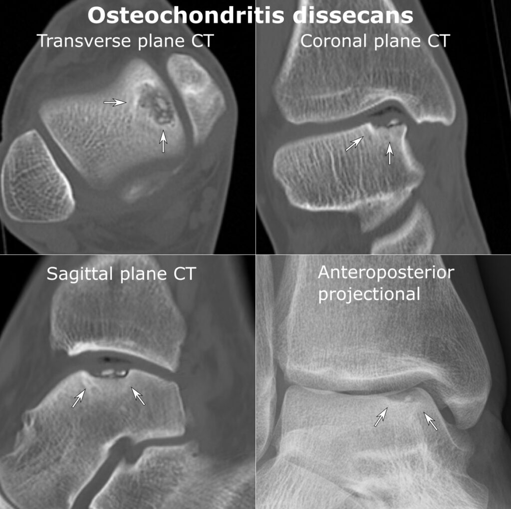
- Radiographs with AP, lateral, tunnel and skyline views
- MRI for size, stability and cartilage integrity
- High T2 signal line or cysts behind the fragment suggest instability
- Hand radiograph for bone age when physeal status is unclear
Classification
- ICRS arthroscopic grading of stability
- Stable lesions with intact cartilage versus unstable or detached fragments
Management
- Stable juvenile lesions, activity restriction for 3 to 6 months with serial imaging
- Persistent stable lesions, transarticular or retroarticular drilling
- Unstable in situ fragments, debridement and fixation with screws or bioabsorbable implants
- Detached salvageable fragments, bed preparation and fixation
- Unsalvageable fragments, removal with osteochondral autograft, allograft or ACI
Prognosis
- Juvenile lesions heal better than adult lesions
- Large lesions and unstable fragments predict poorer outcome
- Untreated unstable lesions predispose to early osteoarthritis
Reviewed by Professor Phong Tran, Head of Orthopaedic Surgery, Western Health. Last updated 10 October 2026.