
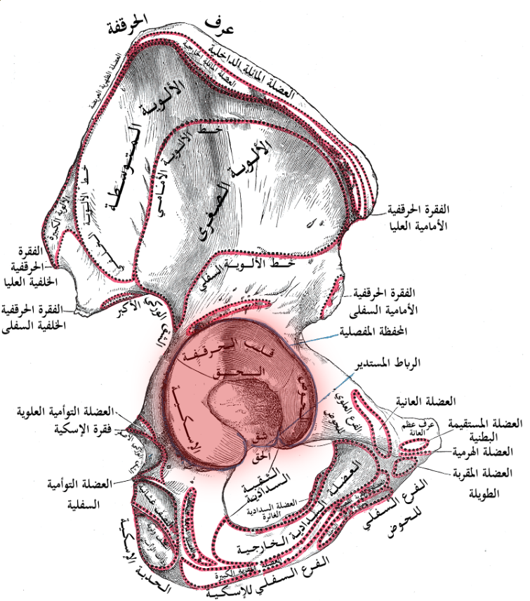
Aims
- To expose the posterior aspect of the acetabulum
Indications
- Posterior lip & column acetabular fractures
- Simple transverse fractures
Position
- Lateral
- If necessary skeletal traction can be used
- Skeletal pin through distal femur
- For Transverse fractures
- Use prone position unless strong assistant
- In lateral position gravity causes medialization of femoral head
Landmarks
- Greater trochanter
- Iliac crest
Incision
- Longitudinal incision centred over greater trochanter
- Start: just below iliac crest
- End: 10 cm below tip of greater trochanter
Internervous Plane
- Gluteus maximus split
Superficial Dissection
- Deepen the incision through subcutaneous fat
- Incise fascia lata in line of skin incision in lower half of wound
- Extend this incision superiorly along the anterior border of gluteus maximus
- Being careful to preserve inferior gluteal nerve
- Retract the split edges to reveal the piriformis muscle & short ER
- Palpate for sciatic nerve
- IR the leg to put the short ER on stretch
- Detach these muscles from insertion into femur
- Leave quadratus femoris intact to protect underlying ascending branch of medial femoral circumflex artery
- Exposes hip joint capsule
- Often torn in trauma
Deep Dissection
- Posterior lip fractures can now be visualised & fixed
- Identify Greater sciatic notch
- For further exposure of posterior column
- 1. Elevate gluteus medius & minimus subperiosteally from posterior & lateral ilium
- 2. Perform an osteotomy of greater trochanter
- Divide GT from posterior to anterior removing a piece of bone 5 cm in size
- Elevate this bone & attached gluteal muscles anteriorly
Dangers
- Nerves
- Sciatic
- Inferior gluteal nerve
- Vessels
- Inferior & superior gluteal artery
- Heterotopic ossification
- Increased with acetabular fractures & trochanteric osteotomy
Reviewed by Professor Phong Tran, Head of Orthopaedic Surgery, Western Health. Last updated 10 October 2026.