
Definition
- Slow growing malignant tumour of the jaw & long bones
- Probable epithelial origin
Epidemiology
- Rare
- 15-30 years of age
- M>F
- Of long bones 90% in diaphysis of anterior tibia
- Rest in fibula or forearm bones
Clinical Features
- Insidious pain onset
- Over many years
- Intracortical stage IA sarcoma
- 15% of patients get metastasis
- 60% had history of trauma to limb months to years prior
Investigations
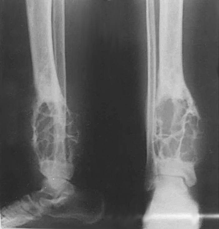
X-ray
- Extensive Soap Bubble appearance
- Multiloculated & cystic lesion
- Surrounding sclerosis, cortical thinning & expansion
- May only involve anterior cortex initially
- Similar to Ossifying fibroma
MRI
- Low intensity T1 signal
- Brighter on T2 that is not diminished with fat suppression
Pathology
Gross
- Well-circumscribed & rubbery nature
- Greyish-white colour
- Focal areas of haemorrhage & necrosis present
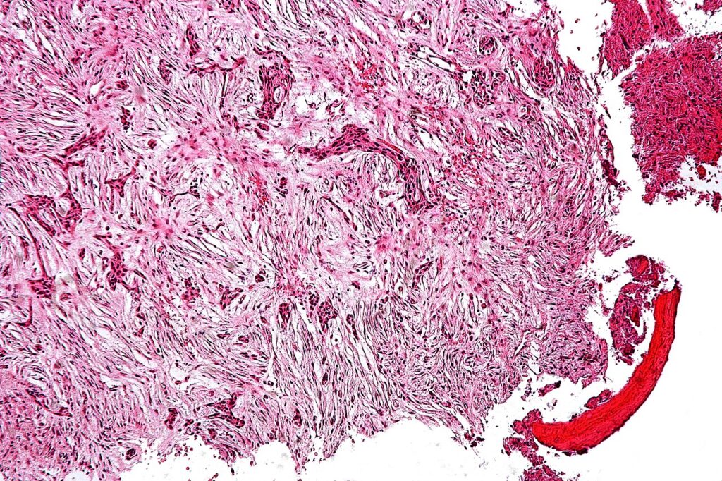
Microscopic
- Biphasic appearance of
- Spindle shaped collagen-producing cells
- Alternating with sinewy cords or nests of epithelioid cells
- Spindle shaped cells probably reactive change only
- Cells similar to fibrous dysplasia
- Positive for Keratin
Treatment
- Wide excision
- Can have satellite lesions some distance from main mass
- Locally invasive & 20% metastasis late in disease
Reviewed by Professor Phong Tran, Head of Orthopaedic Surgery, Western Health. Last updated 10 October 2026.