
Aetiology
- Marfan syndrome is autosomal dominant, caused by FBN1 mutations on chromosome 15
- Abnormal fibrillin weakens connective tissue
- Diagnosis by revised Ghent criteria
Epidemiology
- Scoliosis in 55 to 63% of patients
- Higher rates of thoracolumbar kyphosis and spondylolisthesis
Clinical Features
- Tall stature, arachnodactyly and joint laxity
- Pectus deformity
- Ectopia lentis
- Aortic root dilatation and mitral valve prolapse
- Curves follow idiopathic patterns, often double major
- Thoracic lordosis and thoracolumbar kyphosis
- Curves progress in adulthood
- Assess for protrusio acetabuli and pes planovalgus
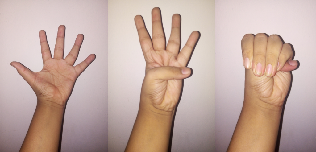
- Joint laxity with positive wrist and thumb signs
Investigations
- Full length standing radiographs
- MRI for dural ectasia before surgery
- Echocardiography and cardiology review before surgery
Management
- Observation for small curves
- Bracing may be tried for moderate curves in growing children
- Bracing less effective than in idiopathic scoliosis
- Most skeletally immature patients with significant curves come to surgery
- Posterior fusion, often with longer constructs than idiopathic curves
- Anterior and posterior surgery for marked kyphosis
Complications
- Dural ectasia with thin pedicles and laminae, increasing screw misplacement and CSF leak
- Higher pseudarthrosis and implant failure rates
- Junctional kyphosis
- Higher infection rates than idiopathic scoliosis
- Cardiovascular risk during anaesthesia, including aortic dissection
- Pneumothorax risk from apical blebs
Differential Diagnosis
- Homocystinuria, with lens dislocating downward and intellectual disability
- Ehlers-Danlos syndrome
- Loeys-Dietz syndrome
- Congenital contractural arachnodactyly
Reviewed by Professor Phong Tran, Head of Orthopaedic Surgery, Western Health. Last updated 10 October 2026.