
Epidemiology
- Scoliosis is the most common skeletal manifestation of neurofibromatosis type 1
- Spinal deformity affects a substantial proportion of patients
Classification
| Type | Features |
|---|---|
| Dystrophic | Short and sharp, involving 4 to 6 vertebrae in the thoracic spine. Often kyphoscoliotic and progress rapidly |
| Nondystrophic | Behave like idiopathic curves |
- Modulation describes a nondystrophic curve becoming dystrophic, mostly in young children
Pathology
- Vertebral scalloping and wedging
- Rib pencilling
- Spindling of transverse processes
- Enlarged neural foramina
- Dural ectasia and intraspinal neurofibromas
- Defective pedicles
Clinical Features
- Café au lait spots, axillary freckling and cutaneous neurofibromas
- Associated cervical kyphosis and spinal instability
- Neurological deficit from intraspinal tumour or severe kyphosis
- Rapid progression or a short sharp curve should prompt assessment for dystrophic change
Investigations
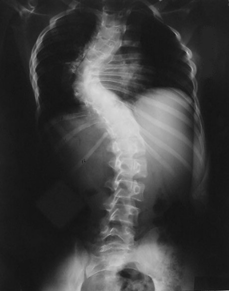
- Full length standing radiographs
- MRI before surgery to detect dural ectasia and intraspinal neurofibromas
- Cervical spine radiographs before anaesthesia and positioning
Management
- Nondystrophic curves treated like idiopathic scoliosis, with closer surveillance for modulation
- Dystrophic curves respond poorly to bracing
- Dystrophic curves need early fusion
- Anterior and posterior fusion when kyphosis is marked
- Growing constructs for early onset dystrophic curves
- Long fusions with bone graft to reduce pseudarthrosis
- Postoperative CT or radiographs to detect pseudarthrosis
Complications
- Higher pseudarthrosis rate than idiopathic scoliosis
- Curve progression after fusion and crankshaft
- Pedicle and screw failure through dysplastic bone
- Neurological injury from dural ectasia or unrecognised tumour
- Haemorrhage from vascular neurofibromas
Reviewed by Professor Phong Tran, Head of Orthopaedic Surgery, Western Health. Last updated 10 October 2026.