
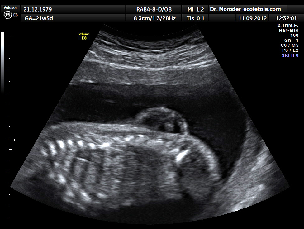
Definition
- Neural tube defects can be grouped under the following terms
- Spina bifida
- Congenital spinal disorder where the two halves of posterior arches fail to fuse leading to bony abnormality
- Spinal dysraphism
- Failure of the neural tube to close
- Myelodysplasia
- Any developmental defect of the spinal cord
- Classification
- 1.Spina Bifida Cystica
- Vertebral laminae absent with prolapse of neural elements/ meninges
- Usually applies to meningomyelocoele
- Meningocoele
- Prolapse of the meninges only
- No neurological deficit
- Meningomyelocoele
- Prolapse of the cord & the meninges in defect
- Neurological deficit present
- Myelocoele
- AKA Myeloschisis, Rachischisis
- Vertebral arches absent & neural tube is unfolded
- Skin & sac absent – most severe form of defect
- Neurological deficit present
- Lipomeningocoele
- AKA Leptomyelolipoma
- Meningocoele that includes a lipoma involving the sacral nerve roots
- 2. Spina Bifida Occulta
- Defect in vertebral arch
- Contained meninges & cord
- No relation to neural tube defects
- “ Occulta “ = Secret
- Seen in 5-10% of radiographs in normal population
- May see telltale skin defects
- Skin dimpling
- Sacral pit
- Hair tuft
- Lipoma
- May have diastematomyelia
- Fibrous, cartilaginous, or osseous bar creating longitudinal cleft in spinal cord
- Can lead to tethering of the cord
- Epidemiology
- 1-4/1000 live births
- Varies with geographic location
- Especially. seen in Celts
- F > M
- Increased to 1/20 with one affected sibling
- Increased to 1/10 with second affected sibling
- Aetiology
- Multifactorial nature
- Genetic & Environmental
- Polygenic inheritance with teratogenic environmental factors
- Decreased RBC folate associated with ↑ risk
•Pathogenesis
- Early problem 24-28th gestational day
- Two theories
- 1. Failure of the neural tube to close
- 2. Initially closed tube that reopens due to ↑ intraluminal pressure
- The latter theory is favoured & explains other defects such as diastematomyelia etc
- Prenatal Screening
- Maternal Alphafetoprotein ↑
- Identifies women at high risk
- Will justify other investigations including
- Amniocentesis
- Detailed USS
•Clinical features
- Dependent on the neurosegmental level (see later)
- CNS Problems
- Spasticity
- 25% of lower limbs
- Causes
- Contracture & deformity
- Difficulty with orthotic fittings
- Impaired walking & sitting
- Poor personal hygiene skills
- More admissions & operations
- Can be treated with
- Intraspinal rhizotomy
- Distal cordotomy
- Deterioration
- Due to other CNS abnormalities
- May be sudden or insidious
- Manifested by
- Increased weakness
- Increased spasticity
- Increased pain
- Discrepancy of > 2 levels between bony anomaly on XR & clinical level
- Rapidly progressive scoliosis
- Decrease of hand function or IQ
- Vocal cord paralysis
- Regular assessment required to detect these
- Detailed neurological examination at each visit
- CNS imaging indicated if deterioration
- Head CT or MRI
•Associated CNS Conditions
- 1. Hydrocephalus
- 90%
- Causes
- Poor IQ
- Poor hand-eye coordination
- Poor fine motor skills
- Most need V-P shunt inserted at time of the defect closure
- Shunt malfunction not uncommon
- Manifested by
- Vomiting/ Nystagmus/ Headache
- Decreased conscious level
- Decreased motor function
- Increased paralysis
- Increased scoliosis
- 2. Hydrosyringomyelia
- 50%
- Related to hydrocephalus
- 4th ventricle communicates with the central canal of cord
- Increased hydrocephalus pushes fluid into cord
- Leads to
- Increased lower limb paralysis & back pain
- Weak upper limbs
- Progressive scoliosis
- Usually settles with the V-P shunt replacement for hydrocephalus
- May need drainage prior to spinal procedure
- Undrained syrinx may cause neurological deterioration
- with curve correction
- 3. Arnold-Chiari Malformation
- Type II (non-communicating) in 90%
- Major features of type II are
- Displacement of Cerebellar Tonsils into Cervical Canal
- Distortion of Medulla Oblongata
- Small shallow Posterior Fossa with enlarged Foramen Magnum
- Symptoms are
- Periodic apnoea
- Stridor
- Weak/ absent cry
- Nystagmus
- Upper limb spasm & weakness
- May resolve with shunt
- If not then surgically decompress
- 4. Tethered Cord
- Almost universal to some degree
- Attachment of cord to meningocoele sac prevents normal upward migration of the cord during growth
- Even with release of cord from all attachments at time of closure there is high likelihood of reattachment during healing process
- Only small number have symptoms
- Buttock & posterior thigh pain
- Increased spasticity & weakness in lower limbs
- Progressive scoliosis
- Treated with surgical release
- Usually arrests progress
- Not restore function
- Neurosegmental Level
- Named according to lowest functioning level
- L4 is key level as quadriceps function » ambulation
•Ambulation Categories (Hoffer)
| Category | Description |
|---|---|
| Community ambulator | Walks indoors and outdoors with or without orthoses, wheelchair only for long trips or speed |
| Household ambulator | Walks only indoors with orthoses, independent or minimally assisted transfers, wheelchair outdoors |
| Non-functional ambulator | Walks only as therapy, wheelchair for mobility |
| Non-ambulator | Wheelchair bound, often able to transfer from bed to chair |
Original publication Hoffer MM, Feiwell E, Perry R, Perry J, Bonnett C. Functional ambulation in patients with myelomeningocele. J Bone Joint Surg Am. 1973;55(1):137-48.
•Classification of Neurological level
- Modified Asher & Olsen
- Tx no grade 3 strength in LL
- L1,2 hip flexion or adduction
- L3 knee extension
- L4 knee flexion
- L5 ankle DF
- Symptoms ankle PF
- Motor testing for neurologic level
- Hip flexion L1, L2, L3 } essentially » flexion & adduction L2,3
- extension L5, S1 } » extension & abduction L4,5
- adduction L2, L3 }
- abduction L4, L5, S1 }
- Knee extension L3, L4
- flexion L5, S1
- Ankle DF L4, L5
- PF S1, S2
- inversion L4
- eversion L5, S1
- Toe DF L5, S1
- PF S1, S2
•Summary for neurosegmental level
Summary of neurosegmental level
| Level | Functioning muscles | Mobility |
|---|---|---|
| Thoracic | No voluntary lower limb function | Wheelchair |
| High lumbar (L1 to L3) | Hip flexors, with or without hip adductors and knee extensors | Children use HKAFO or KAFO, 75% of adolescents and adults use a wheelchair |
| Low lumbar (L4, L5) | As above plus knee extensors and flexors and ankle dorsiflexors, with or without hip abductors | 75% of adults are community ambulators, most with an AFO |
| Sacral (S1 to S4) | As above plus ankle plantarflexors, with or without toe flexors | Community ambulators for limited distances, with or without shoe orthoses |
- Specifics for neurosegmental level
- Thoracic Level
- » Non-walkers
- Power
- No voluntary muscle activity in lower limb
- No fixed deformity
- Position
- Legs in position of gravity
- ER at hips
- Slight knee flexion
- Equinus of ankle
- Menelaus
- ? Higher rate of hip dislocation in this group than L2/ 3 levels
- ? Not related to muscle imbalance across hip
- Ambulation
- Child
- Standing frame at 12-18/12 for 2-3 years
- RGO in early years
- Ie. Nonfunctional ambulators
- Helps them to develop more normally & decreases contractures
- Adults
- Wheelchair as too much energy for mobilisation
- Upper Lumbar L1-3
- » Nonambulators 75%
- » Household ambulators 25%
- L1 Level
- Power
- Some hip flexion from Psoas (L1,2,3) =2/5
- Position
- Hip flexed, abducted & ER
- Ambulation
- Child
- Possible to use RGO/ HKAFO
- Adult
- Usually wheelchair-bound
- L2 Level
- Power
- Hip flexion from Psoas =3/5
- Some adduction (L2,3) =3/5
- Position
- Hip flexed & adducted
- FFD of hip may develop
- Ambulation
- Child
- Most ambulate as children in HKAFO
- FFD may need to be corrected first
- Adult
- Usually wheelchair-bound
- L3 Level
- Power
- Hip flexion & adduction =4-5/5
- Quads (L3,4) =3/5
- Position
- Hip flexed & adducted
- Knee extended
- Hip subluxation & dislocation
- Common due to unopposed hip adduction & flexion
- Ambulation
- Usually household ambulators
- If quads are 3/5 then
- need KAFO to walk
- 88% in wheelchair
- If quads are 4/5 then
- 98% household ambulators
- 82% community ambulators
- Quads & no abductors » can ambulate with splint
- Quads & good abductors » can ambulate without aids
- Lower Lumbar L4-5
- » Community ambulators 75%
- L4 Level
- Power
- Quads =5/5
- Hamstrings (L5, S1) =3/5
- Tibialis Anterior (L4) =3-4/5
- Tibialis Posterior (L4,5) =1/5
- Position
- Hip flexed & adducted
- Knee extended
- Ankle in varus
- Foot variable*
- Ambulation
- Walk in AFO
- Surgery often required to maintain
- Hip extended
- Knee extended
- Foot plantigrade
- L5 Level
- Power
- Hip abduction (L4-5, S1) =3-4/5
- Tibialis anterior & posterior =3-4/5
- Position
- Calcaneus foot or Calcaneovalgus*
- Ambulation
- 95% community ambulators throughout life
- Foot surgery often required
- *Low Lumbar with Spastic Sacral Segment
- In addition to L4
- Spastic hip abductors?
- Spastic hamstrings
- Spastic calf
- Spastic peronei
- Deformity
- Equinocavo-Valgus or Equinovalgus (peronei spastic)
- Equinovarus (tib posterior spastic)
- Calcaneus due to weak Achilles
- Vertical Talus
- Club Foot
- Sacral Level
- S1 Level
- Power
- Hip extension (gluteus maximus (L5, S1,2) =3/5
- Gastrocnemius (S1) =3/5
- Toe flexors (S1,2) =4-5/5
- Position
- Pes cavus
- Clawing of toes from intrinsic minus position
- Ambulation
- Usually brace free but may need special shoes
- S2 Level
- Power
- Normal
- Position
- May have claw toes
- Ambulation
- Normal
- muscle charting should start immediately post-op
- 1/3 Complete LMN lesion & loss of sensation & bowel control below affected level
- 1/3 Complete lesion at some level but distal segment of cord preserved with mixed picture of intact DTR & spasticity
- 1/3 Incomplete & some movement & sensation preserved
- Other deformities common such as
- DDH
- CTEV
- Genu recurvatum
- Claw toes
- Initial Treatment
- Selection of patients for closure of the defect is controversial
- Two protocols
- 1. Some centres avoid urgent operation if
- Level above L1
- Severe deformity
- Marked hydrocephalus
- If not treated most die of meningoventriculitis
- Survive if given antibiotics
- More severely handicapped due to
- Hydrocephalus
- Continuing trauma to cord
- 2. Now almost all infants treated initially
- » Ethically difficult to decide to not treat
- Team orientated approach
- Neurosurgery
- Urology
- Orthopaedics
- Physio
- OT
- Initial treatment involves
- Closure of defect within 24 hours
- V-P shunt insertion
- Orthopaedic Management
- Menelaus Principles
- 1. Always manage in Spina Bifida clinic
- 2. Select surgery appropriate to future demands
- 3. Perform minimal surgery
- 4. Condense management
- 5. Correct muscle imbalance
- 6. Consider absent sensation, bone fragility, likelihood of infection
•Principles
- Promote walking if possible to
- Promote normal bony development
- Prevent contractures
- Surgery is soft tissue
- Release contractures
- Tendon transfers
- All surgery at one sitting
- Simple surgery most appropriate in high level lesions
- Wait until 12 months as
- Spinal level & deformities evident & assessable
- Most shunt & spine closure complications have been corrected
•Goals
- General
- Ambulation requires
- Range of motion
- Absence of contracture
- Stability
- Motor power
- Co-ordination
- If surgery considered
- More important to achieve ROM
- Because stability can be provided by orthosis
- Specific
- Mobile & symmetrical hips
- Ambulators ability to extend knees
- Chair-bound ability to flex knees
- Plantigrade foot
- Straight spine
- Indications for surgery
- Posture stable
- Pelvis level
- Personal hygiene
- Pressure sores
- Preserve respiration
•Hips Thoracic Level
- Aim at achieving ROM
- Functional range without contracture required to allow,
- Sitting in wheelchair
- Lying in bed
- Orthoses for standing & walking
- Surgery in form of contracture release
- Reduction of hip not required
- No sensation or muscle control in legs
- Dislocation not common
- If occurs, likely due to CNS problem
- Dislocation usually doesn’t reduce function
- Even if unilateral
- Reduction of dislocation difficult & often fails
- Avoid Multiple Procedures
- Leads to scarring
- Causes loss of functional range
- Orthotics required for lower limb stability
- Must include hip, knee, ankle & foot (HKAFO)
- Most give up walking by 8 years
- Even if orthosis available
- Parapodium, Swivel walker
- Reciprocating Gait Orthosis may be better
- Use wheelchair because
- Uses less energy
- Quicker
•Hips Upper Lumbar
- High incidence of hip dislocation
- Unopposed hip flexion & adduction
- Attempts to reduce hip often difficult
- Due to uncorrected muscle imbalance
- Best treatment is to release contractures causing FFD & adduction
- Release of Iliopsoas & Adductors
- Release Rectus Femoris if tight
- Then treat as a thoracic level
- Better to have flaccid flexible hip than a strong stiff hip
•Hips Middle / Lower Lumbar
- Hip usually undergoes progressive dysplasia & dislocation
- Due to hip flexion & adduction during stance phase for stability
- Progressive acetabular dysplasia
- Dislocate age 3-4
- Unilateral disease in child with good quads & potential walker should be reduced
- Traditional treatment was muscle release
- But may lead to weakness & ↑ instability
- Requires orthosis to stabilise hip for walking
- Another option is tendon transfer
- Iliopsoas to Greater Trochanter (Sharrad transfer)
- Decreased hip dysplasia
- Problems with stair climbing
- Weakness of hip flexion means Rectus must be used to flex hip
- Causes knee extension & difficulty climbing stairs
- Can be remedied with physiotherapy
- Hip reduction must be performed first
- At age 1 year
- Open reduction
- Varus shortening femoral osteotomy performed for femoral neck deformity
- Pelvic osteotomy performed for acetabular dysplasia
- Reduction of Hip (Menelaus)
- No Quads & bilateral » Never } above L4
- No Quads & unilateral » Sometimes }
- Quads & Bilateral » Sometimes } L4 & below
- Quads & Unilateral » Always }
•Knees
- Aim for straight knee that is braceable
- Straight knee is stable position
- Quadriceps weakness
- Treatment with KAFO
- Extension contracture
- May interfere with walking
- Most can be treated with serial casts & splints
- If resistant, VY-plasty of quadriceps tendon
- Flexion contracture
- Not important in chair-bound
- Treatment in ambulators by
- Release Hamstring & Gastrocnemius
- ± Posterior capsulotomy
- If near skeletal maturity with large FFD could consider distal femoral extension osteotomy
•Ankles & Feet
- Rigid & flail deformities in anaesthetic feet
- Majority have Deformity
- 50% have Equinovarus deformity
- 20% have Calcaneus deformity
- 20% have Normal feet
- 10% have Valgus, Equinovalgus, Cavus & Claw Toe deformities
- Aim For Braceable Plantigrade Feet
- Almost all will require brace
- Equinovarus
- Most common deformity
- Due to lesion at L3 or L4
- Splintage & casting initially
- Well padded serial casting as insensate skin
- Stretching often corrects varus
- May require percutaneous lengthening of TA
- Attempt casting until age 6 months
- If fails, operate at age 12 months
- Usually PMR
- If fails, may require talectomy
- If fails, triple arthrodesis after skeletal maturity
- Calcaneus
- L5 level most common
- Due to unopposed
- Tibialis anterior
- Toe extensors or peronei
- And weak
- Gastrocnemius
- Os calcis becomes vertical
- Leads to heel ulceration
- Treatment with release of offending tendons
- Leave treatment until 3 years to fully assess problem
- ? Dynamic EMG to assess tendon transfer
- Tibialis anterior to calcaneus
- Valgus
- Usually less problem than varus
- Caused by
- Lateral tilt of ankle mortise
- Valgus subtalar joint
- Can be managed often with AFO
- Can be treated by
- Supramalleolar osteotomy
- Grice subtalar arthrodesis
- Transfer tends to fail
- Cavus
- Sacral level
- Weak Tendo Achilles
- ± Claw toes
- Due to lack of intrinsic power S2
- Corrected by
- Jones Tendon Suspension
- Tendon transfer EHL to metatarsal heads & IPJ fusion
- Steindler stripping
- Tarsal/ Metatarsal osteotomy
•Spine
- Scoliosis
- Most common skeletal deformity (80%)
- More common with high lesion
- Aetiology
- May be due to 3 factors
- Congenital
- Due to congenital malformations
- Neuromuscular
- Combination of
- Paralysis
- Instability with no posterior elements
- Neurological
- Progressive neurological abnormality
- Hydrosyringomyelia
- Failed VP shunt
- Tethered cord
- Curve similar to idiopathic curve
- Management
- Orthotics
- Temporary measure
- To delay fusion
- In order to allow development of trunk height
- Problems with pressure sores
- Surgery
- Need good dorsal soft tissue
- Indications for intervention
- Failure of orthotic management to maintain curve < 45°
- Scoliosis in 12 yo previously controlled with brace
- Progressive spinal decompensation
- Progressive pelvic obliquity
- In the form of
- Anterior release & fusion + Posterior segmental fusion
- Avoids Crankshaft effect
- If > 12 yo consider posterior only
- Kyphosis
- Difficult problem
- Trouble sitting
- Ulcerations over kyphus
- Can’t see ahead
- Breathing difficulties
- May be treated with
- Excision of Kyphotic segment
- Excision of distal cord
- Lordosis
- Usually corrected by correction of FFD of hip
- Difficulties with spinal surgery
- Deficient posterior elements
- Previous sac repair
- Anterior fusion forms the basis
- Blood loss higher
- Infection rates higher
•Foot & Ankle 2
- Lateral tilt of ankle in mortise
- May require supramalleolar osteotomy with medial closing wedge if severe enough
- If sufficient growth remaining then consider medial epiphysiodesis of the distal tibial physis (< 6 years of age)
- Subtalar
- Peroneal tendon releases
- If fails then consider Grice ST fusion & calcaneal osteotomy
- If close to maturity then look at Triple Arthrodesis
- Cavus
- Pressure effects major problem
- Up to 5 years consider Plantar release
- If fails then look at Metatarsal osteotomies
- If associated varus of heel then add calcaneal osteotomy
- If close to maturity & significant deformity then look at Triple Arthrodesis
- Claw toes treated with Jones procedure & Hibbs to lesser toes & PIPJ fusion
- Extensor tenotomies & dorsal capsulotomies of MTPJ
- CVT
- < 2% of children with SB
- Similar reduction as per other forms of CVT
- Should attempt in first year of life
- External Rotation of Tibia
- Commonly associated with valgus ankle
- Requires supramalleolar osteotomy > 8 yo if severe
•Fractures
- Most often around knee
- Most common at age 3-7 years
- Often painless
- Only diagnosed with
- Redness
- Warmth
- Swelling
- Usually heal with abundant callus/ heterotopic ossification
- Usually treat nonoperatively
- Avoid disuse if possible
•Urinary Complications
- Major cause morbid/ mortality
- Spastic paralysis of bladder
- Leads to
- Urinary incontinence
- Hydronephrosis
- Recurrent UTIs
- Renal failure
- Addressed by
- Intermittent catheterization
- Bladder augmentation
•Latex Allergy
- Latex is an organic substance obtained from rubber tree
- Ubiquitous in hospitals, households, community
- Sensitisation occurs during surgical procedures
- Incidence of latex reactions related to number of previous operations
- Can develop
- Contact dermatitis type IV hypersensitivity)
- Urticaria, rhinoconjunctivitis, asthma, anaphylaxis (type I hypersensitivity)
- Fatalities reported in the US
Reviewed by Professor Phong Tran, Head of Orthopaedic Surgery, Western Health. Last updated 10 October 2026.