
Definition
- Avascular necrosis & subsequent disintegration of lunate
- Described 1910 by Kienbock (Vienese radiologist)
Aetiology
- Avascular necrosis of lunate
- Probably result of trauma
- Mechanical & vascular theories
Vascular Theory
- Due to trauma disrupting vascularity
- May be
- Single incident with disruption of blood supply
- Multiple compression fractures with loss of blood supply to fragments
- Lunate vascularity (Gelberman dye studies)
- 8% Single incomplete palmar feeder
- Hence higher risk AVN
- Severe hyperextension may disrupt it
- 92% Dorsal & Palmar blood vessels
- “X” “I” “Y” configuration
- Well-vascularised
- Need intra & extraosseous disruption
- Low risk AVN
- 8% Single incomplete palmar feeder
Mechanical Theory
- Ulnar variance
- Normal +3 to -6 mm (2 SD mean)
- Ulnar minus variance
- Subjects lunate to greater compression & shear forces
- More common in Kienbock’s
- Gelberman 1975
- 75% with Kienbock’s cf. 25% in unaffected patients
- Assess on standard XR
- Realise that true Kienbock’s rarely seen as result of perilunate/ lunate dislocations (<1%)
Epidemiology
- Occurs in young active adults
- Age 20-40
- Usually dominant hand
- Rarely bilateral
Anatomy
- Lunate articulates with radius, scaphoid, triquetrum, capitate and, in type 2 lunates, hamate
- Mostly covered in cartilage, with vessel entry limited to dorsal and volar surfaces
- Gelberman intraosseous patterns Y, I and X, with the I pattern possibly at higher risk
- Negative ulnar variance associated with Kienbock disease (Hultén)
Pathology
- Avascular necrosis of the lunate with progressive collapse
- Sclerosis, then fragmentation and loss of lunate height
- Proximal migration of the capitate and fixed scaphoid flexion
- Carpal collapse leading to radiocarpal and midcarpal arthritis
- Articular cartilage status graded arthroscopically (Bain)
Classification
Lichtmann
Classified into 4 stages according to X-ray
| Stage | Findings |
|---|---|
| 1 Normal | No radiographic changes. May be possible to see on MRI |
| 2 Sclerosis | Density changes. Sclerosis of proximal pole |
| 3A Fragmentation | May be evidence of compression fracture. Radiolucent or radiodense line |
| 3B Collapse | Collapse of lunate. Flattening |
| 4 Degeneration | Pancarpal arthritis |
Original publication Lichtman DM, Mack GR, MacDonald RI, Gunther SF, Wilson JN. Kienböck’s disease: the role of silicone replacement arthroplasty. J Bone Joint Surg Am. 1977;59(7):899-908.
- Operations aimed at promoting revascularisation for stages 1 & 2
History
- Gradual onset of pain & stiffness in wrist
- Usually no history of trauma (but seen in up to 50%)
Examination
- Tenderness over lunate
- Passive dorsiflexion MF pain
- Diminished grip strength
- In later stages ↓ ROM from Osteoarthritis
Investigations
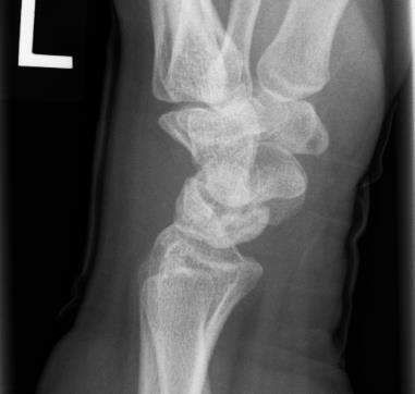
Plain X-ray
- Progressive changes of avascular necrosis
- Mottling
- Collapse
- Degenerative arthritis
- XR for Ulna Variance
- AP film with wrist in neutral
- Elbow flexed 90°
- Shoulder abducted 90°
Bone Scan
- Increased uptake in early stages
MRI
- Shows changes before bone scan becomes positive
Differential Diagnosis
- Ulnar impaction syndrome
- Intraosseous ganglion of the lunate
- Acute lunate fracture
- Preiser disease
- Inflammatory arthritis
- SLAC or SNAC wrist
Treatment
Non-operative
- No treatment is satisfactory way of managing Kienbock’s
- Beckenbaugh 1980 70% no worse at 7 years
- Kristensen 1986 77% minimal pain at 18 years
- Saffar 1982 few changed work at 10-30 years
Splintage
- ? Allows fractures to heal & revascularise
- Rarely effective
Operative
Early – Stage 1, 2 & 3
- Ulna Minus
- Radial shortening
- Rationale is to redistribute stresses
- Decreased stress on lunate may allow it to revascularise
- Try to achieve ulnar neutral wrist
- ~ 2mm shortening
- 2mm = 20% ↓ in radial load
- Volar T Plate
- Good-excellent results 80-90% success
- Can consider combining with vascularised graft into lunate using metacarpal artery
- Ulnar lengthening
- Other form of joint levelling
- Don’t do as high rates of nonunion
- Radial shortening
- Neutral Ulna Variance
- Radial closing wedge
- Ulna opening wedge
- Capitate-Hamate Fusion
- Arthroscopic drilling (forage)
Late – Stage 4 (? 3 Also)
- Limited fusion
- Scaphotrapeziotrapezoid (STT) fusion
- Unloads lunate fossa & transfers load to scaphoid fossa
- Unpredictable
- Silicone replacement
- Poor long-term results
- No longer advocated
- Proximal row carpectomy
- ? Best procedure for late cases
- Can consider Buck-Gramko denervation if pain is main symptom
Operative Salvage Procedures
- Arthrodesis of wrist
- Manual laborers
- Proximal Row Carpectomy
- Good if Low Demand
- Lunate Excision
- Controversial
- Good results in 1 study (Kawai)
Management Approach
- Literature not proven to show operative approach better than observation alone
- Therefore observation is reasonable option for all stages of Kienbocks
- Stage 1
- Observe
- Stage 2
- Observe
- Arthroscopic drilling (forage)
- Radial osteotomy ± vascularised bone graft
- Stage 3
- Observe
- Radial osteotomy ± VBG
- If carpal collapse then scapholunate capsulorraphy
- Stage 4
- Observe
- Radial osteotomy
- Denervation
- Total fusion
Complications
- Progressive collapse and wrist arthritis
- Nonunion of radial shortening osteotomy
- Ulnar wrist and DRUJ pain after radial shortening
- Hardware irritation
- Radiocapitate arthritis after proximal row carpectomy
- Stiffness and loss of motion after partial or total wrist fusion
- CRPS
Prognosis
- STT fusion or Radial Shortening doesn’t prevent collapse
Reviewed by Professor Phong Tran, Head of Orthopaedic Surgery, Western Health. Last updated 10 October 2026.