
Carpal instability is loss of normal alignment and motion between the carpal bones, most often from scapholunate ligament injury causing dorsal intercalated segment instability (DISI). Untreated instability can progress to scapholunate advanced collapse (SLAC) arthritis.
Definition
Situation where normal alignment of carpal bones is lost
Aetiology
- DISI
- due to disruption of scapho-lunate articulation
- VISI
- secondary to disruption of lunate & triquetral
- Ulnar translocation
- rarely results from injury but is commonly seen in wrists affected by rheumatoid arthritis
Epidemiology
- Scapholunate dissociation is the most common carpal instability
- Lunotriquetral instability is less common
- Young adults after falls and sporting injuries
- Scapholunate injury is often associated with distal radius fractures
- Frequently missed at initial presentation
Anatomy
- Eight carpal bones arranged in proximal and distal rows
- Proximal row is an intercalated segment with no tendon insertions
- Scaphoid links proximal and distal rows
- Under load the scaphoid tends to flex and the triquetrum to extend
- SL and LT ligaments balance these opposing forces through the lunate
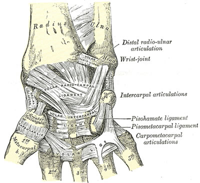
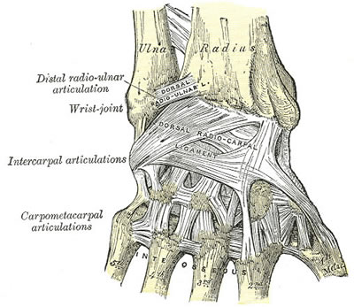
- Two major groups of ligaments
- extrinsic ligaments course between carpal bones & radius or metacarpals
- intrinsic ligaments originate & insert in carpus
- extrinsic ligaments are stiffer while intrinsic ligaments are capable of greater elongation before permanent deformation occurs
Extrinsic
Palmar
- palmar extrinsic ligaments consist of 2 V-shaped ligamentous bands
- one is proximal & connects forearm to proximal carpal row
- proximal limb consists of
- radiolunotriquetral & radioscaphoid ligs laterally
- ulnolunate & ulnotriquetral ligs medially
- proximal limb consists of
- one is distal & connects forearm to distal carpal row
- distal limb of palmar extrinsic ligs consists of
- radioscaphocapitate ligament laterally
- ulnocapitate ligament medially
- distal limb of palmar extrinsic ligs consists of
- one is proximal & connects forearm to proximal carpal row
Dorsal
- dorsal extrinsic ligaments
- radiotriquetral
- scaphotriquetral
Intrinsic
- intrinsic ligs connect adjacent carpal bones
- Proximal row
- intraarticular
- connecting scaphoid to lunate & lunate to triquetrum
- strong
- critical for carpal stability
- intraarticular
TFCC
- separates ulna from carpus
- Made up of
- TFC
- dorsal & volar radio-ulna ligaments
- ulnar collateral ligament
- meniscus homologue
- articular disc
- sheath of ECU
- Variable thickness of TFC from 1 – 5mm depending on station of ulna (ulna variance)
- No tendons are directly attached to carpus
- Made up of
Rows/Columns
- Traditional view
- proximal & distal row of carpal bones- scaphoid joining them
- Taliesnik
- three columns
- scaphoid being radial column
- triquetral ulnar column
- lunate & capitate with remaining carpal bones making up middle column
- three columns
- Navarro
- Three column modification
- scaphoid, trapezium & trapezoid making up radial column
- triquetral ulnar column
- capitate & lunate along with hamate middle column
- Three column modification
Pathology
- Ligament disruption causes loss of normal carpal kinematics
- SL disruption lets the scaphoid flex and the lunate extend, producing DISI
- LT disruption lets the lunate flex with the scaphoid, producing VISI
- Untreated SL dissociation progresses to SLAC arthritis
- SLAC begins at the radial styloid, then radioscaphoid, then capitolunate, sparing radiolunate
- Perilunate injuries progress from radial to ulnar (Mayfield stages)
Classification
4 Major Types of Carpal Instability
| Type | Description |
|---|---|
| DISI | Dorsi-flexion (Dorsal Intercalated Segment Instability). Most common, where lunate is rotated into dorsi-flexion (zig zag alignment of radiolunatocapitate alignment) |
| VISI | Palmar flexion (Volar Intercalated Segment Instability) |
| Ulnar translocation | Abnormal translocation of lunate ulnarward Type 1 entire carpus is translocated ulnarward Type 2 relationship between radius & scaphoid is normal but scapholunate gap is wide Rheumatoid |
| Dorsal subluxation | Malunion fracture distal radius with reversal of normal palmar tilt |
Original publication Linscheid RL, Dobyns JH, Beabout JW, Bryan RS. Traumatic instability of the wrist. Diagnosis, classification, and pathomechanics. J Bone Joint Surg Am. 1972;54(8):1612-32.
Instabilities May Be
| Type | Description |
|---|---|
| Static | Loss of normal alignment can be seen on XR |
| Dynamic | Routine XR within normal limits. Instability can be produced by either voluntary movement or manipulation, eg between scaphoid + lunate, between lunate + triquetrum, or at midcarpal joint |
Instabilities May Also Be Termed
| Type | Description |
|---|---|
| Dissociative | S-L dissociation DISI L-T dissociation VISI |
| Nondissociative | May also result in VISI or DISI but 3 bones (S, L, T) act as unit, eg dorsal carpal subluxation, mid-carpal instability, Type 1 ulnar translocations |
History
- Fall on outstretched hand with wrist extension and ulnar deviation
- Dorsal wrist pain and weak grip
- Painful clicking or clunking with loading
- Previous wrist injury and duration of symptoms
- Hand dominance and occupation
Examination
- painful wrist
- clicking or clunking
- Ballottement test
- Watson test for scaphoid instability
Investigations
- AP
- AP wrist under axial load (clenched fist)
- hand in radial & ulnar deviation
- Findings
- May demonstrate distance between scaphoid & lunate or lunate & triquetral
- DISI pattern
- scapholunate gap
- ring sign
- with flexed scaphoid seen end on
- scaphoid foreshortened
- distance between ring & proximal pole less than 7mm
- flexed scaphoid is seen with dorsiflexed lunate
- (quadrilateral) & with triquetrum in distal (dorsiflexed) position
- VISI pattern
- ring sign
- scaphoid foreshortened
- lunate volar flexed (triangular)
- triquetrum distal in relation to hamate (dorsiflexed)
- distance between ulnar head & triquetrum is reduced (Mayersbach sign)
- convex outline of proximal carpal row (= Shentons line of wrist) is interrupted by step off between lunate & triquetrum
- Ulnar Translocn
- Carpal-Ulnar distance
- is distance from centre of head of capitate (ie centre of rotation of carpus) & line produced along line of centre of ulna
- Normally ratio of C-U distance/ length of 3rd metacarpal = .30 ± .03
- ¯ in ulnar translocation
- Carpal-Ulnar distance
- Lat: to assess opposite rotations of scaphoid & lunate
- DISI pattern
- when scapholunate joint is dissociated
- scaphoid is palmar flexed
- lunate is dorsiflexed
- Scapho-lunate angle
- Normal 30- 60° (av 46o)
- DISI > 70°
- when scapholunate joint is dissociated
- VISI pattern
- lunate palmar flexed
- lunotriquetral angle
- Normal -16 deg
- Abn neutral or +ve
- Ulnar Translocation
- often associated with VISI
- SLAC wrist (scapho-lunate advanced collapse)
- With S-L dissociation
- All load going through Radioscaphoid joint
- degenerative process
- radial styloid & scaphoid
- luno-capitate joint (commonest pattern of degeneration 55%)
- triscaphoid degeneration
- between scaphoid, trapezium + trapezoid
- 2nd most common pattern
- With S-L dissociation
- Other Ixs
- Bone scan
- is useful to identify pathology
- When bone scan is negative it suggests either that there is no injury or more frequently that problem is minor & can be treated non operatively
- Arthrography
- is helpful in finding ligament tears but? significance as these may not necessarily be result of trauma but may indicate age related degenerative change
- NB: need to compare with normal side
- Arthroscopy
- can directly visualise pathology
- Bone scan
Differential Diagnosis
- Scaphoid fracture or non union
- TFCC tear
- Dorsal wrist ganglion
- Kienböck disease
- Midcarpal instability
- Radiocarpal or STT arthritis
- ECU subluxation
Treatment
- of chronic instability depends on patients symptoms
- Nonsurgical
- little disability
- > 80% of ROM
- > 80% grip strength
Scapholunate dissociation
- Acute
- either closed manipulation or open reduction with pinning
- Chronic: if no associated Osteoarthritis
- reattachment of scapholunate ligament
- dorsal capsuloligamentodesis (Blatt)
- dorsal capsular flap used to prevent scaphoid from subluxing in palmar direction
- ref: Blatt & Nathan ” Dorsal Capsulodesis for rotatory subluxation of scaphoid: review of long term results” Proc Am Soc of Surgery of Hand 1992
- STT fusion
- problem is with radial impingement
- S-L or S-C fusion
- if associated Osteoarthritis (ie SLAC)
- Excise (?replace) scaphoid & perform mid carpal fusion that is fusion of capitate, hamate, triquetral & lunate
- (= 4 – corner fusion)
- total wrist fusion
Lunatotriquetral instability
- Acute
- either immobilisation in BEPOP or direct repair of ligament
- chronic
- Lunatotriquetral arthrodesis
Ulnar Translocation
- Acute
- Repair of disrupted volar & dorsal radiocarpal ligs
- Chronic
- ligament repair unreliable
- relocation of carpus & maintenance of reduction by radiolunate arthrodesis more reliable
Dynamic VISI & DISI
- trial of nonoperative management with AEPOP/ NSAIDS/ local injection of steroids
- Stabilisation of midcarpal joint limited fusion
- Capsuloligamentodesis
- tenodesis
Complications
- SLAC wrist
- Persistent pain and stiffness after reconstruction
- Failure of repair with recurrent instability
- Loss of wrist flexion after capsulodesis
- Superficial radial nerve injury and pin site infection
Prognosis
- Acute repair within about six weeks gives better results than late reconstruction
- Static instability has a worse prognosis than dynamic instability
- Untreated SL dissociation progresses to SLAC wrist
- Salvage procedures relieve pain at the cost of motion
Reviewed by Professor Phong Tran, Head of Orthopaedic Surgery, Western Health. Last updated 10 October 2026.