
Tibial shaft fractures are the most common long bone fracture and are often open because the medial tibia is subcutaneous. Intramedullary nailing is the standard treatment for displaced fractures, and compartment syndrome must be watched for after both injury and surgery.
Epidemiology
- 1/2000 per annum
- Commonest long bone fracture
- Fractures of the proximal third make up 5-10% of fractures in most series
Classification
- Most surgeons use descriptive classifications
- Can use AO classification
- degree of soft tissue injury can be classified by the system of Tscherne & Gotz (1984)
| Type | Description |
|---|---|
| 0 | ~Minimal soft tissue damage resulting from an indirect mechanism of injury that has caused a simple bone fracture |
| 1 | ~Superficial abrasion or soft tissue contusion ~caused by pressure from the bone injury with a mild to moderately severe fracture pattern |
| 2 | ~Deep contaminated abrasion ~associated with localised skin & muscle contusion, an impending compartment syndrome & a high energy fracture pattern |
| 3 | ~Extensive skin contusion or crushing ~underlying severe muscle damage, a compartment syndrome & a severe fracture pattern |
Compartment Syndrome
- rate of compartment syndrome varies from 1-9%
- rate of compartment syndrome is no higher when reaming is used compared with no reaming
- compartment pressure is most elevated by the use of continual traction rather than intramedullary reaming
Indications for Nonoperative or Operative Treatment
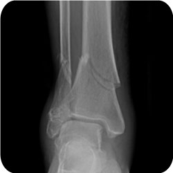
Distal Tibial & Fibular Shaft Fracture
- Published alignment parameters are guidelines at best, with no substantiated scientific data to support them
- Some accepted guidelines are:
- Varus/valgus angulation of 5-7°
- AP angulation of 10°
- Shortening of 1cm
- Rotational alignment within 10°
- There is no consensus on the best management of a closed midshaft stable tibia fracture amongst trauma experts
- In a meta-analysis by Littenberg et al (JBJSA 1998) the only strong conclusions that could be reached were that closed treatment has a lower risk of infection & open treatment has a higher rate of union
- presence of an intact fibula leads to more rapid union but is associated with an ↑ risk of angulatory deformity
Displaced Tibial Fractures
- 1991 RCT by Hooper reported that treatment of displaced tibial fractures by IMN resulted in a better outcome than closed treatment
- with more rapid union
- less malunion
- earlier return to work.
- Advantages & disadvantages of closed vs. open treatment
Closed Treatment
- Negligible risk of infection
- Few problems with knee pain
- No need for hardware removal
Intramedullary Nailing
- Better control of alignment
- Can start early ROM of knee & ankle
- Improved mobility
- Less frequent followup
- Earlier return to work
Present Indications for Nonoperative Management of Tibial Fractures
- Minimal soft tissue injuries (types 0 & 1 by Tscherne & Gotz)
- Stable fracture pattern:
- less than° coronal angulation
- less than 10° sagittal angulation
- less than 1cm of shortening
- Ability to bear weight in a cast or functional brace
Indications for Nailing
- High energy fracture
- Types 2 & 3 soft tissue injuries
- Unstable fracture pattern by above definitions
- An open fracture
- Compartment syndrome
- Ipsilateral femoral fracture
- Inability to maintain reduction
- Intact fibula (relative indication)
Points on Nailing
- Reaming is preferred to non-reaming.
- A larger, stiffer nail can be used which leads to less hardware breakage
- less risk of non union & repeat operations
- Proximal tibial fractures have a much higher rate of complications than midshaft fractures
- rate of nonunion is up to 84% compared with 34% in midshaft fractures
- Malunion occurs as a result of malreduction
- fracture tends to collapse into valgus
- due to loss of more lateral cortex than medial cortex
- attachment of the anterior tibial muscles on the lateral cortex acting as a tether
- fracture tends to posteriorly translate (particularly if the fracture is proximal to the bend in the nail) & flex
- fracture tends to collapse into valgus
- To avoid malreduction the entry point should be anterior & laterally
- Tornetta found the ideal entry point is 3mm lateral to the midpoint of the tibial tubercle
- Blocking (Poller) screws & unicortical plating are techniques that can be used to ↓ the risk of malunion
- Poller screws are placed posteriorly & laterally
- Flexion deformity was minimised by Tornetta by using a small medial arthrotomy with a semi-extended position
- Distal tibial fractures
- have less of a tendency to malunion than proximal fractures but are still more problematic than proximal fractures
- Technical points here
- percutaneous clamps can be used to maintain a reduction
- Plating the fibula may ↓ the rate of malunion
- A Steinmann pin placed horizontal to the joint line acts as a visual aid to reduction & can be used as a joy stick
Nailing & Open Fractures
- Reaming has traditionally been thought to be contraindicated in open tibial fractures because of damage to the endosteal blood supply
- Two recent RCT have shown no ↑ in the rate of infection with reaming
Complications of Nailing
- Knee pain
- occurs in around 50%
- Not influenced by patellar splitting or medial parapatellar approach
- Abolished by nail removal in 50% & ↓ in 25%
- Nailing leads to ↑ patellofemoral contact forces
- Nonunion
- Bone grafting is safe after 3 months in grade 3A or 3B fractures if there is no evidence of infection
- Fibular nonunion may also occur & be a source of pain. This needs to be treated with bone grafting & compression plating
- Malunion – 12-34%
- Delayed union
- Consider prophylactic bone grafting at 6 weeks if using small diameter unreamed nail
- Rule out infection at the time of Reoperation
- Hardware failure
- This is reduced if a large reamed nail is used
- Two distal locking screws should be used – one study reported a rate of screw failure of 59% with a single screw vs. 5% with two screws
Plating
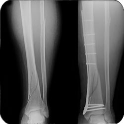
Spiral Distal Tibial & midshaft Fibular Shaft Fracture treated with Medial Locking Plate
- Plating is used mainly for metaphyseal injuries. It should not be used when there is soft tissue compromise
- plate can be placed laterally (fewer soft tissue problems & biomechanically more advantageous because acts as a tension band) or medially (preferred if subcutaneous placement)
External Fixation
- External fixation may be definitive or provisional
- Provisional external fixation is the treatment of choice in injuries where there is dubious viability of the limb
- Definitive external fixation is reserved for patients with:
- very narrow intramedullary canals (less than 6mm)
- children
- patients with complex periarticular fractures
- All studies comparing Ex-fix & nonreamed nails in managing open fractures have found better results for nailing.
- rates of deformity were lower
- faster return to weight bearing
- improved limb function
- use of IMN simplified soft tissue cover & bone grafting operations
- Typically the frame is placed anteromedially
- with four pins
- two close to the fracture but not within the fracture haematoma
- other pins as far distal as possible
- connecting rods are initially placed as close to the skin as possible
- but some dynamization can be achieved by moving the rods further away from the skin
- with four pins
- Complication
- pin loosening & subsequent pin tract infection
- If the Ex-fix is to be replaced by an IMN
- pin tracts should be healed, as numerous authors have documented an ↑ rate of infection if nailing is performed after more than 2 weeks of Ex-fix
- Predrilling of all pin sites should be performed, as this may ↓ the rate of thermal necrosis
Results
| Procedure | Time to Union (weeks) | Non / delayed / Union | Malunion | Superficial Infection | Deep Infection | Reoperation |
|---|---|---|---|---|---|---|
| Closed Treatment | 17.2% 13.1% delay 4.1% non | 31.7% | 0% | 1/145 | 12/145 | |
| ORIF | 14.9 weeks | 2.6% 0.86% delay 1.7% non | 0 | 9.0% | 1/233 | 11/233 |
| Unreamed Nail | 19.5 | 16.7% 9.4% delay 7.4% non | 11.8% | 0.5% | 3/203 | 31/203 |
| Reamed Nail | 20.2 | 8.0% | 3.2% | 2.9% | 3/314 | 19/314 |
Reviewed by Professor Phong Tran, Head of Orthopaedic Surgery, Western Health. Last updated 10 October 2026.