
Sternoclavicular joint dislocations are uncommon and usually follow high energy trauma. Anterior dislocations are more common and often managed without surgery, while posterior dislocations can compress the trachea, oesophagus or great vessels and need urgent reduction with a cardiothoracic surgeon available.
Definition
- Dislocation of the Sternoclavicular Joint
Incidence
- Extremely uncommon injury
Aetiology
- most commonly injured by MVA’s, then sports
- caused by lateral compression injury to arm
Anatomy
- Stability of SC joint
- is provided by joint capsule/costoclavicular & interclavicular ligaments
- very little joint congruity
- Medial epiphysis fuses with clavicle at 23-25 years; therefore, injuries in young adults often physeal
Classification
| Basis | Types |
|---|---|
| Direction | Anterior, more common by far Posterior, more serious injury, least common, can present with venous engorgement, SOB, difficulty swallowing secondary to obstruction of mediastinum |
| Mechanism | Traumatic, atraumatic |
| Degree | Dislocation, subluxation |
- Spontaneous Atraumatic Subluxation
Examination
- Diagnosis can be difficult on physical examination
Investigations
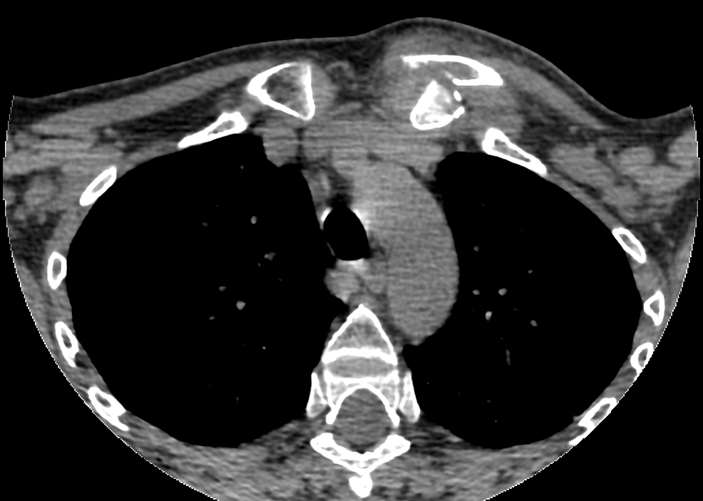
X-rays
- radiographs often are nondiagnostic
- AP & 40° cephalic tilt view
CT Scan
- most reliable scan to show subluxation / dislocation
- shows relation also to vascular structures & associated fractures
Treatment
All Subluxations & Sprains
- treat nonoperative
Anterior Dislocation
- usually managed nonoperatively with activity modification & reassurance
- may do closed reduction
- traction & abduction
- bump between shoulders
- often unsuccessful
- many remain unstable & usually not improved by open intervention
- figure 8 splint for 6 weeks
- persistent prominence is usually present but not of functional significance
– atraumatic dislocation
– no specific treatment is required, as the natural history is relatively good
– traumatic dislocation: some patients may have pain & loss of function
Posterior Dislocation
- may require treatment because of proximity of major neurovascular structures & airway
- closed reduction
- thorough vascular exam preop including CT scan to assess vascular injury & proximity
- under GA in operating room
- chest / vascular / cardiothoracic surgeon available to address any potential vascular or airway catastrophe associated with injuries to the mediastinum
- Use abduction & traction & towel clip
- Successful closed reduction usually stable
- avoid Internal fixation because of likelihood of hardware migration & possible injury to the mediastinal structures
- If closed reduction unsuccessful, open reduction is indicated
Physeal Injuries
- should generally be left alone to remodel
- if symptomatic & posterior
- then closed reduction can be done
- Posterior dislocations require pre-op CT to assess vascular injury, & these require CR under GA with vascular notification
Spontaneous Subluxator or Dislocator
- Do not operate
Recurrent or Irreducible Posterior Dislocations in Adults
- may require medial clavicle excision & costoclavicular ligament reconstruction
Osteoarthritis from Chronic Dislocation
- may resect SCJ (Operative Technique – SC Joint Excision)
Complications
- Potential Vascular Injury
- Bump (cosmetic)
- Degenerative Joint Disease
- Mediastinal Impingement with Posterior Dislocation
Prognosis
- Recurrent instability uncommon
- Many apparent dislocations in adolescents may be growth plate injuries that will remodel without treatment
Reviewed by Professor Phong Tran, Head of Orthopaedic Surgery, Western Health. Last updated 10 October 2026.