
Moore
Southern
Principles
- do no interfere with abductor mechanism of hip
- avoid loss of abductor power
Position
- lateral position
- protect & pad bony prominences
- prep & drape limb free
Landmarks
- Greater Trochanter
- Iliac crest
- Shaft of femur
Incision
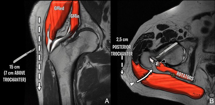
- 15 cm curved incision centred on the posterior aspect of greater trochanter
- Start: begins 8 cm above & posterior to the posterior aspect of the greater trochanter
- End: down shaft of femur
Internervous Plane
- No true internervous plane
- Gluteus maximus split
Superficial Dissection
- Incise fascia lata on lateral aspect of femur to uncover vastus lateralis
- Lengthen the fascial incision superiorly in line with skin incision & split the fibres of gluteus maximus by blunt dissection
- Ligate branches of superior & inferior gluteal arteries that are encountered
- Retract fibres of gluteus maximus
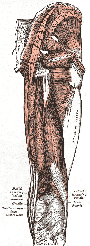
- Exposes short external rotators & overlying fatty tissue
- Obturator internus
- Two gemelli
- Quadratus femoris
Deep Dissection
- Palpate & identify the sciatic nerve
- IR the hip to put the short IR on stretch & put the operative field farther from the sciatic nerve
- Stay sutures into piriformis & obturator internus tendons
- Detach the muscles close to their femoral insertion
- Reflect them backwards to cover & protect the sciatic nerve
- If necessary I would extend my dissection to include quadratus femoris
- But haemostasis of the branches from the lateral circumflex artery can be problematic
- Exposes hip joint capsule
- Place stay suture in capsule
- Incise with a T shaped capsulotomy
Dangers
- Nerves
- Sciatic
- Vessels
- Inferior gluteal artery
- Lateral circumflex artery
Reviewed by Professor Phong Tran, Head of Orthopaedic Surgery, Western Health. Last updated 10 October 2026.