
Definition
- CPPD deposition disease is calcium pyrophosphate crystal deposition in articular and periarticular tissues
- Chondrocalcinosis is radiographic calcification of hyaline cartilage or fibrocartilage
Classification
- Asymptomatic deposition
- Osteoarthritis with CPPD
- Acute CPP crystal arthritis (pseudogout)
- Chronic CPP inflammatory arthritis
Epidemiology
- Prevalence rises steeply with age, uncommon before 60 unless metabolic or familial
- Onset before about 55 years prompts a metabolic screen
Aetiology
- Excess chondrocyte pyrophosphate combines with calcium to form crystals
- Familial forms involve ANKH, which regulates pyrophosphate transport
- Shed crystals activate the NLRP3 inflammasome, releasing interleukin 1 beta
- Haemochromatosis, with hook-like osteophytes on the second and third metacarpal heads
- Hyperparathyroidism and hypomagnesaemia, including Gitelman syndrome
- Hypophosphatasia, with low alkaline phosphatase
- Osteoarthritis, previous joint trauma and meniscectomy
Clinical Features
- Acute hot swollen joint, sometimes with fever, mimicking septic arthritis
- Triggered by surgery (especially parathyroidectomy), illness, trauma and intra-articular hyaluronic acid
- Chronic arthritis in atypical joints, radiocarpal, glenohumeral, patellofemoral and MCP
- Crowned dens syndrome, acute neck pain and fever from odontoid calcification
Investigations
- Aspiration shows weakly positively birefringent rhomboid crystals, blue when parallel to the compensator
- Gout crystals are strongly negatively birefringent needles
- Always send Gram stain and culture, as infection can coexist
- Metabolic screen of calcium, PTH, magnesium, phosphate, ALP, iron studies and thyroid function
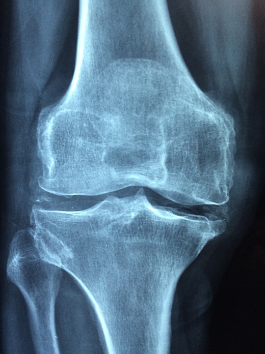
- Chondrocalcinosis in knee menisci, triangular fibrocartilage and pubic symphysis
- Isolated patellofemoral arthritis, large subchondral cysts and SLAC wrist
- Ultrasound shows a hyperechoic band within hyaline cartilage
Differential Diagnosis
- Septic arthritis and gout
- Rheumatoid arthritis and inflammatory osteoarthritis flare
- Basic calcium phosphate (hydroxyapatite) arthropathy
Management
- Acute attacks treated with aspiration, rest, ice and NSAIDs or colchicine
- Intra-articular corticosteroid once infection is excluded
- Systemic corticosteroid for polyarticular attacks or when other agents are contraindicated
- Interleukin 1 inhibitors for refractory disease
- Treat metabolic causes, although chondrocalcinosis does not regress
- No equivalent of urate-lowering therapy
- Low dose colchicine prophylaxis, magnesium for hypomagnesaemia
- Arthroplasty for end-stage arthritis
- Postoperative crystal flares mimic infection, so aspirate and culture
Reviewed by Professor Phong Tran, Head of Orthopaedic Surgery, Western Health. Last updated 10 October 2026.