
Definition
- Calcium Pyrophosphate Dihydrate Deposition Disease
- Chondrocalcinosis can refer to any calcium in cartilage
- CPPD
- CaPO4
- HA
Classification
| Type | Notes |
|---|---|
| Idiopathic chondrocalcinosis | By far the most common. Pseudorheumatoid arthritis with continuous attack, pseudo osteoarthritis with acute attacks, pseudo osteoarthritis without acute attacks, lanthanic (asymptomatic) |
| Hereditary | |
| Secondary (“WHIP A DOG”) | Wilson’s, haemochromatosis, hyperparathyroidism, hypothyroidism, hypophosphatasia, idiopathic, pseudogout, pernicious anaemia, acromegaly, diabetes, degenerate, ochronosis (alkaptonuria), gout |
Pathology
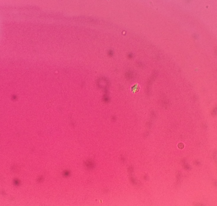
- Chondrocalcinosis is the finding of radioopaque crystals in hyaline cartilage & can be salts of
- Calcium pyrophosphate dihydrate
- Dicalcium phosphate hydrate
- Hydroxyapatite
- Calcium oxalate
- May have acute or chronic presentation
- Joints involved
- Knee
- Hip without symptoms often
- Patellofemoral
- Radiocarpal
- Metacarpophalangeal
- Joints involved
Investigations
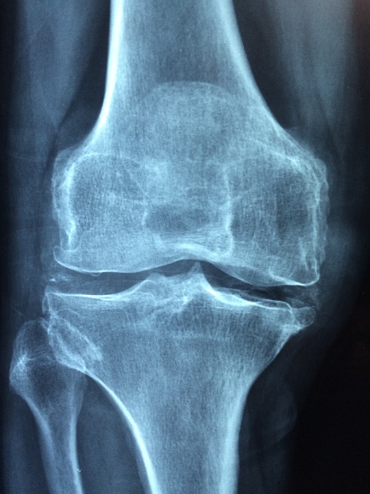
X-rays
- Intraarticular crystals
- Cartilage loss
- Subchondral cysts
- Structural collapse of the articular surface
Differential Diagnoses
- Hyperparathyroidism
- XR show subperiosteal erosions
- Blood tests show hypercalcaemia & ↑ PTH
- Haemochromatosis
- Characteristic
- Progressive degenerative arthritis of finger joints
- XR shows calcification of multiple joints & discs
- Serum iron & iron binding capacity are raised
- Characteristic
- Adult Onset Hypophosphatasia
- Hereditary metabolic disturbance
- Characterised by low levels of serum alkaline phosphatase
- Ochronosis / Alkaptonuria
- Inborn error of metabolism
- Tyrosine pathway
- Homogentisic acid oxidase is absent
- Homogentisic acid accumulates
- Homogentisic acid deposited as a dark brown pigment in
- Connective tissue
- Hyaline & Fibrocartilage
- Presents in 4th decade
- Pain & stiffness
- In spine & larger joints
- XR shows calcification of intervertebral discs
- Progressing to obliteration of the disc space
- And bony ankylosis
- Excretion of homogentisic acid
- Causes the urine to turn dark on standing
Epidemiology
- M:F – 2:1
- Usually patient > 50 years
- Sometimes familial
- Associated with
- DM
- Hypothyroidism
- Gout
- Hyperparathyroidism
- Haemochromatosis
- Pernicious Anaemia
- Ochronosis
Pathology
- CPPD crystals deposited in
- Joint capsule
- Articular cartilage
- Fibrocartilage (Menisci)
- Histologically
- Crystals seen at margin of degenerate cartilage
- Surrounding lacunae of chondrocytes
Pathogenesis
- Pyrophosphate generated at chondrocyte surface in abnormal cartilage
- By enzyme action
- Combine with calcium to form crystals which “sit” in the cartilage
- ? Alter biomechanical properties
- Occasionally the crystals are released into the joint & arthritis results
- Activation of vasoactive & chemotactic factors
- Neutrophils attracted & phagocytose crystals
- Release of lysosomal enzymes into joint fluid
- Crystal release caused by
- Sudden change in ionic Calcium & Pyrophosphate
- Equilibrium in cartilage
- Eg. after acute illness or surgery
- Physical disruption of cartilage
- Eg. due to trauma
- Enzymatic degradation of matrix
- Eg. in inflammatory arthritis
Chronic Chondrocalcinosis
- Predisposes to development of secondary osteoarthritis
- Crystals embedded in articular cartilage have desiccating effect
Clinical Features
- 3 Different Conditions
Asymptomatic Chondrocalcinosis
- Majority of cases
- Common incidental finding is calcium of menisci
Pseudogout
- Usually affects large joints
- Knee > Shoulder > Wrist
- Cf. Gout affects small joints
- Usually monoarticular
- Less pain than Gout
- Rapid onset with peak in 24/24
- Subsides in 1/52
- May be provoked by
- Trauma
- Surgery
- Illness
Chronic CPPD Arthropathy
- Pseudo-Osteoarthritis
- Usual features of Osteoarthritis
- Polyarticular diseases like Osteoarthritis
- Ie. Hips & knees
- ? Due to CPPD in cartilage altering the biomechanics
- In more unusual joints for Osteoarthritis
- Eg. Ankles, shoulders, elbows
- Key to differentiating from normal Osteoarthritis
- Pseudo-Rheumatoid Arthritis
- Acute synovitis & chronic arthritis
- Rapidly progressive joint destruction
Investigations
- Laboratory Tests
- Synovial fluid
- CPPD crystals seen extracellularly & in neutrophils
- Rhomboidal
- Weakly positive birefringent
- CPPD crystals seen extracellularly & in neutrophils
- Ie. Blue parallel to 1st order red filter & 135° to polarizer
- WCC ~ 1000 x 10.6 /L
- Especially neutrophils
- Synovial fluid
- Radiology
- Calcium in articular cartilage
- Fine linear densities
- Parallel to subchondral bone
- Calcium in fibrocartilage & connective tissue
- More punctate densities
- Common fibrocartilage with calcium deposits
- Menisci
- TFCC
- Pubis
- Annulus
- Findings of Osteoarthritis usually present
- Involves unusual sites such as
- NWB Joints
- PFJ
- TNJ
- Calcium in articular cartilage
Differential Diagnosis
- Gout
- Septic arthritis
- Osteoarthritis
- RA
- Spondyloarthropathy
Management
- Symptomatic only
- Crystals deposition can’t be prevented or reversed
- Treatment consists of NSAID
- May use intra-articular corticosteroids
- Joint washout if severe
Reviewed by Professor Phong Tran, Head of Orthopaedic Surgery, Western Health. Last updated 10 October 2026.