
Posteromedial Approach to Ankle

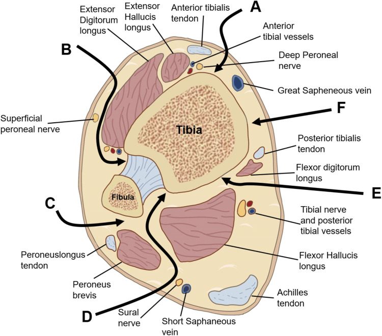
Cross section of the distal leg showing the intervals used to reach the distal tibia, including the posteromedial corridor between the poste
Cross section of the distal leg showing the intervals used to reach the distal tibia, including the posteromedial corridor between the posterior tibial tendons and the tibial neurovascular bundle. Image from Liu J, Zhang Z, Qu J, Piao C. Progress of fracture mapping technology based on CT three-dimensional reconstruction. Front Bioeng Biotechnol. 2024, 12, 1471470. CC BY 4.0.
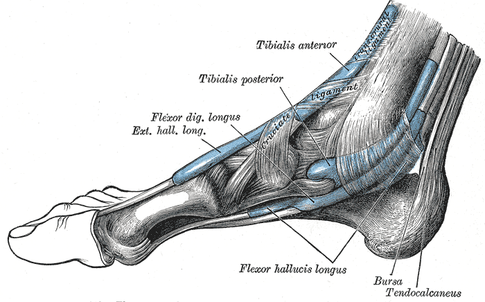
Posteromedial Approach to Ankle, tendon sheaths around the ankle, medial aspect, gray’s anatomy plate 442
Tendon sheaths around the ankle, medial aspect, Gray’s Anatomy plate 442. Image by Henry Vandyke Carter, Wikimedia Commons, Public domain.

Indications

  • club foot
  • exploration of soft tissues around back of malleolus

Position

  • supine
  • leg flexed & externally rotated to expose medial side of ankle
  • tilt table

Landmarks

  • medial malleolus
  • achilles tendon

Incision

  • 10 cm longitudinal incision midway between medial malleolus & achilles tendon

Superficial Dissection

  • subcutaneous fat
  • fascia
  • identify achilles tendon

Deep Dissection

  • 3 ways to approach back of ankle
    • between FHL & peroneal tendons
    • between FHL & FDL
    • between medial malleolus & tibialis posterior & FDL

Dangers

  • Nerves
    • Posterior tibial artery & nerve
  • Vessels

Reviewed by Professor Phong Tran, Head of Orthopaedic Surgery, Western Health. Last updated 10 October 2026.