
Position
- supine
- tourniquet
- prep & drape
Landmarks
- medial malleolus
Incision
- 10 cm curved longitudinal incision centred over the tip of the medial malleolus
Internervous Plane
- None
- Dissection straight down to bone
Superficial Dissection
- Mobilise the skin flaps
- Taking care not to damage the
- Long saphenous vein
- Saphenous nerve
- Taking care not to damage the
Deep Dissection
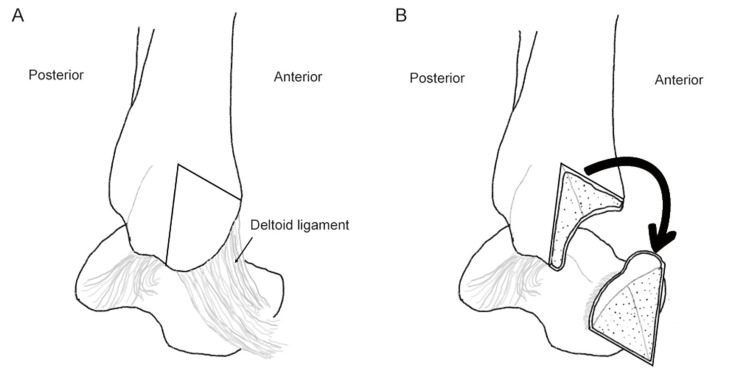
- Osteotomy
- Identify the point at which the MM joins the shaft of tibia
- Small longitudinal incision in anterior part of joint capsule
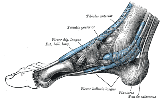
- Divide the flexor retinaculum
- Identify the tendon of tibialis posterior & retract it posterior to expose the posterior surface of malleolus
- Score longitudinally to ensure correct alignment of malleolus during closure
- Pre-Drill & tap
- Osteotome & oscillating saw
- Cut through medial malleolus obliquely from top to bottom
- Checking the position via the anterior arthrotomy
- Retract medial malleolus with attached deltoid ligament downwards
- Forcibly evert the foot exposing the dome of talus & articulating joint surface of tibia
Dangers
- Nerves
- Saphenous nerve
- Vessels
- Tendon of tibialis posterior
Reviewed by Professor Phong Tran, Head of Orthopaedic Surgery, Western Health. Last updated 10 October 2026.