
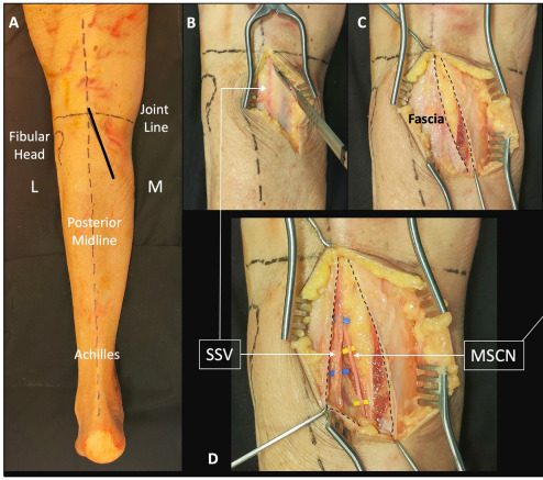
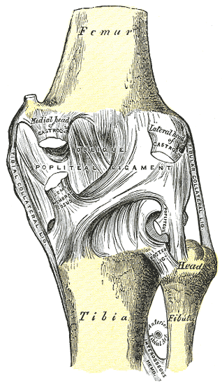
Aims
- Primarily a neurovascular approach
Position
- prone
- longitudinal bolsters to support chest & abdomen & allow expansion
Landmarks
- 2 heads of gastrocnemius
- semimembranosus & semitendinosus
- biceps tendons
Incision
- longitudinal gently curved incision
- Start: laterally over the biceps femoris muscle
- Obliquely across the popliteal fossa
- End: over medial head of gastrocnemius & inferior over calf
Superficial Dissection
- reflect the skin flaps
- identify small saphenous vein & medial sural cutaneous nerve on its lateral side (branch of tibial nerve)
- incise the fascia of popliteal fossa medial to small saphenous vein
- trace the medial sural cutaneous nerve proximally back to its source
- dissect up to the apex of the popliteal fossa following the tibial nerve
- dissect the common peroneal nerve at the apex of the popliteal fossa
- it runs along the posterior border of biceps femoris
- identify the popliteal artery & vein
- lies deep & medial to tibial nerve
- artery has 5 branches
- two superior
- two inferior
- one middle genicular artery
- may need to be ligated to mobilise the artery
- Distally
- Vein is medial to artery
- Then it moves posterior to artery
- Then lies posterolateral to artery above the knee joint
Deep Dissection
- Retract the muscles that form the boundaries of popliteal fossa to expose the various parts of posterior joint capsule
- Can detach either medial or lateral heads of gastrocnemius
Dangers
- Nerves
- Medial sural cutaneous nerve
- Tibial nerve
- Common peroneal nerve
- Vessels
- Small saphenous vein
Reviewed by Professor Phong Tran, Head of Orthopaedic Surgery, Western Health. Last updated 10 October 2026.