
Medial Parapatellar Approach to the Knee

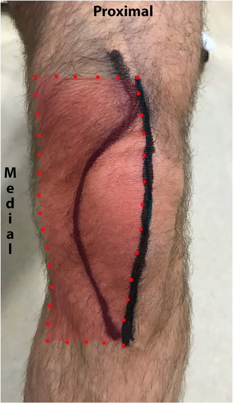
Midline skin incision and the line of the medial parapatellar arthrotomy drawn on a left knee, with the arthrotomy extending proximally into
Midline skin incision and the line of the medial parapatellar arthrotomy drawn on a left knee, with the arthrotomy extending proximally into the quadriceps tendon. Image from Bjørn S, Nielsen TD, Jensen AE, Jessen C, Kolsen-Petersen JA, Moriggl B, Hoermann R, Bendtsen TF. The Anterior Branch of the Medial Femoral Cutaneous Nerve Innervates Cutaneous and Deep Surgical Incisions in Total Knee Arthroplasty. J Clin Med. 2024, 13(11), 3270. CC BY 4.0.
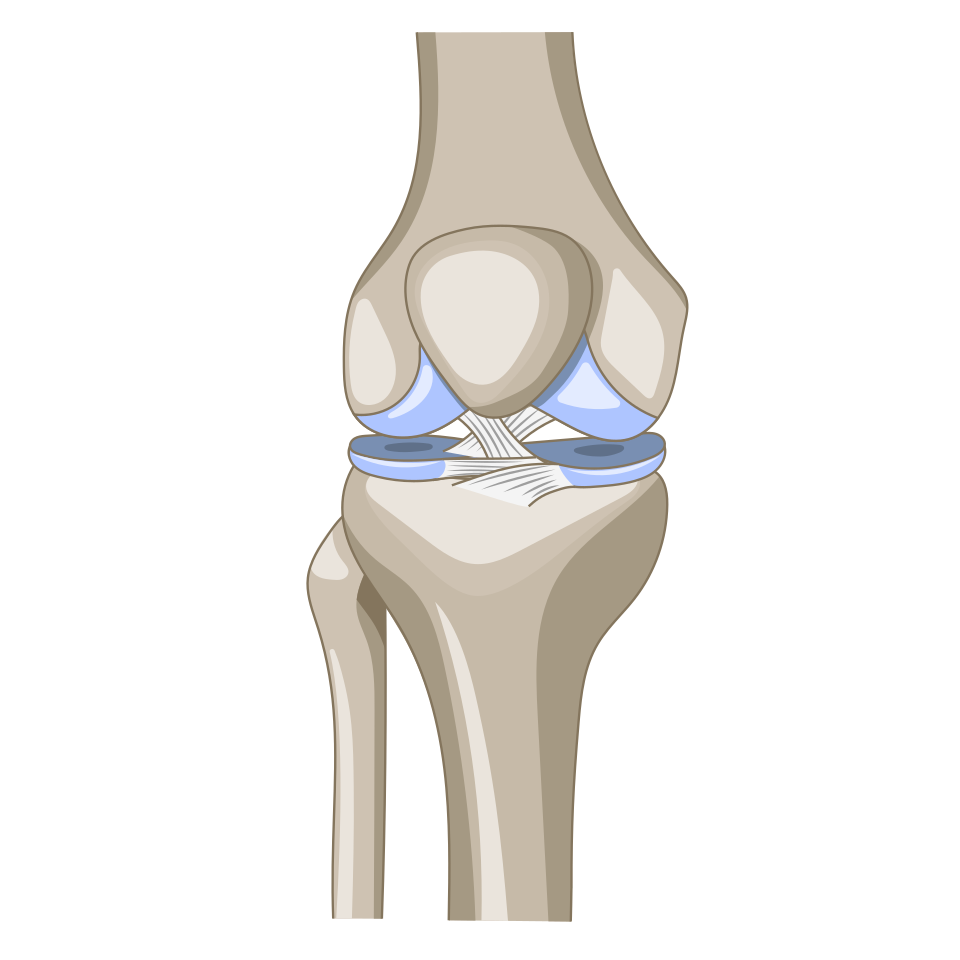
Anterior view of the knee joint
Anterior view of the knee joint. The medial parapatellar arthrotomy runs along the medial border of the patella and patellar tendon. Image by DataBase Center for Life Science (DBCLS), Wikimedia Commons, CC BY 4.0.

Position

  • supine
  • tourniquet
  • foot rest
  • lateral support

Landmarks

  • patella
  • patellar ligament
  • tibial tuberosity

Incision

  • straight longitudinal midline incision extending from a point
  • Start: 5 cm above the superior pole of patella
  • End: level of tibial tubercle

Internervous Plane

Superficial Dissection

  • Subcutaneous tissues
  • Expose quadriceps tendon, medial border of patella, medial border of patella tendon
  • Enter the joint by incising
    • Medial side of patella leaving a cuff for repair
    • Divide quadriceps tendon along its medial border leaving a cuff for repair
    • Dividing the fibrous tissue along the medial aspect of patella tendon
  • Retract the fat pad & excise it

Reviewed by Professor Phong Tran, Head of Orthopaedic Surgery, Western Health. Last updated 10 October 2026.