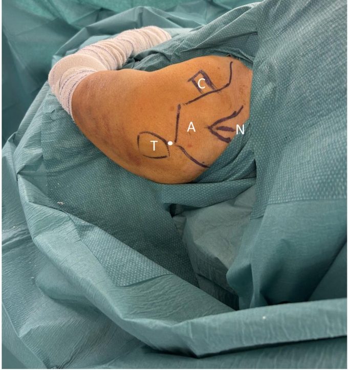
 Surface landmarks for the entry point of an antegrade proximal humeral nail. The white dot marks the K wire entry between the greater tuberosity (T) and the acromion (A), with the coracoid (C) and Neviaser portal (N) also marked. Image from Giovannetti de Sanctis E, Bige B, Cornacchini J, Bronsard N, Gonzalez JF, Gauci MO. Three-part proximal humerus fracture, a full-percutaneous severely displaced greater tuberosity reduction and intramedullary nailing technique. JSES Rev Rep Tech. 2025, 5(4), 1129-1137. CC BY 4.0.
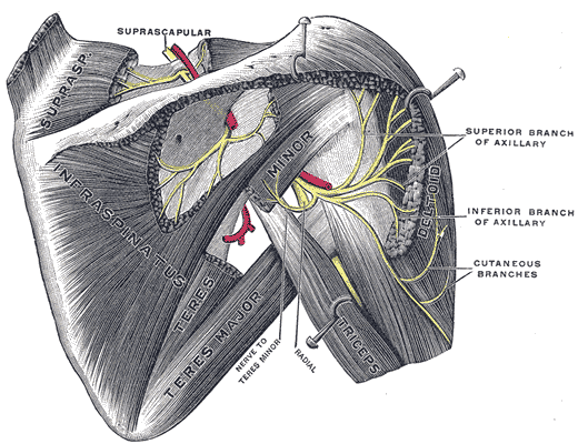 Suprascapular and axillary nerves from behind. The axillary nerve runs about 5 cm below the acromion, which sets the safe zone for the deltoid split and proximal locking screws. Image by Henry Vandyke Carter, Gray’s Anatomy (1918), Wikimedia Commons, public domain.
Indications
- Insertion of humeral nail
Principles
- Entry point
- Lateral to articular surface of humeral head
- Medial to greater tuberosity
Position
- Supine
- Beach chair 60°
- Shoulder is over edge of table
- Mayfield head rest
- Image intensifier
Landmarks
Incision
- 2cm longitudinal incision
- Start: outer aspect of acromion
Internervous Plane
Superficial Dissection
- Insert guide wire under II control
- Cut down onto wire with scalpel through deltoid & supraspinatus
Dangers
Reviewed by Professor Phong Tran, Head of Orthopaedic Surgery, Western Health. Last updated 10 October 2026.
