
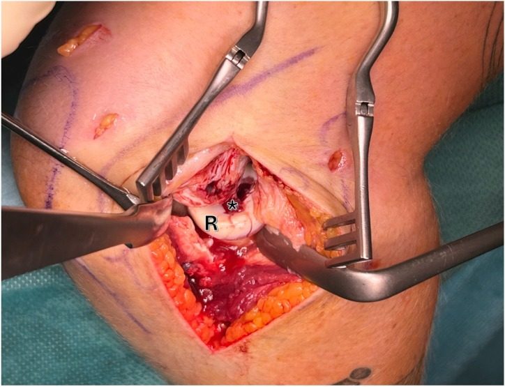
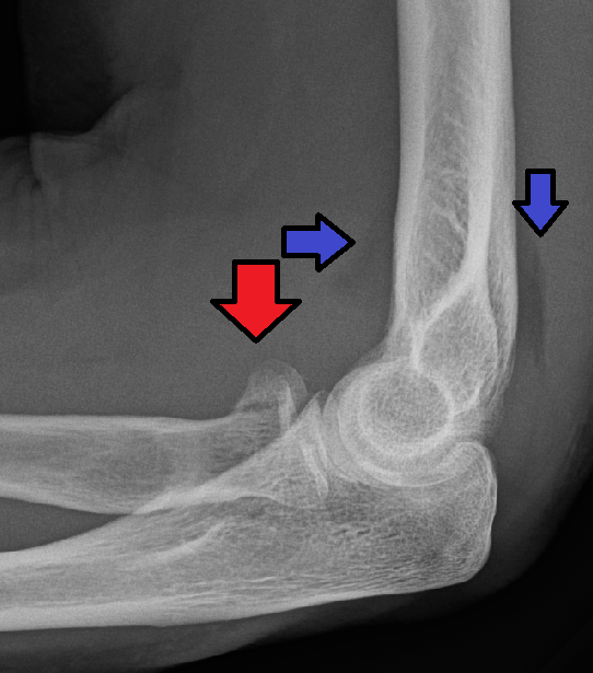
Indications
- ORIF
- Excision
- Replacement
Position
- Supine
- Arm positioned over chest
- Pronate forearm
Landmarks
- Lateral humeral epicondyle
- Radial head
- 2.5 cm distal to lateral epicondyle
- Olecranon
Incision
- 5 cm longitudinal incision
- Start: Posterior surface of lateral humeral epicondyle
- Continued downward & medially
- End: towards the posterior border of ulna
Internervous Plane
- Anconeus — radial nerve
- ECU — PIN
Superficial Dissection
- Deepen skin incision through fat & deep fascia
- Identify the plane between anconeus & ECU
- Develop it with blunt dissection & detach part of the superior origin of anconeus from the lateral humeral epicondyle
Deep Dissection
- Fully Pronate forearm to protect the PIN
- Incise capsule longitudinally
- Capitellum
- Radial head
- Annular ligament
- To expose capitellum
- Cut down onto lateral supracondylar ridge
- Elevate subperiosteally
Dangers
- PIN
Reviewed by Professor Phong Tran, Head of Orthopaedic Surgery, Western Health. Last updated 10 October 2026.