
Indications
- ORIF
- Coronoid process
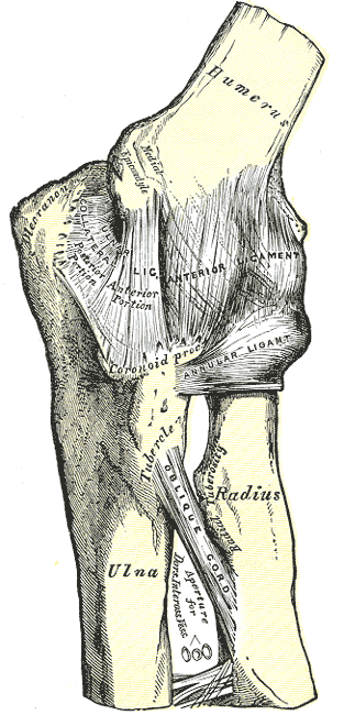
- Medial condyle & epicondyle
Contraindications
- Exploration of elbow
- Access to lateral side
Position
- Supine
- Arm board
- Shoulder fully ER
- Elbow flexed to 90°
- Prep & drape arm free
Landmarks
- Medial epicondyle
Incision
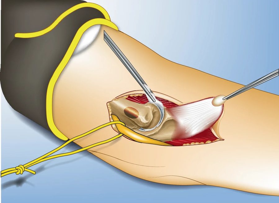
- 10 cm longitudinal incision centred over medial epicondyle
Internervous Plane
- Proximally
- Brachialis
- Triceps
- Distally
- Brachialis
- Musculocutaneous nerve
- Pronator teres
- Brachialis
Superficial Dissection
- Locate ulnar nerve
- Mobilise & hold free with vascular tape
- Identify & develop the plane between pronator teres & brachialis
- Being careful not to damage the median nerve anteriorly
- Gently retract pronator teres medially
Deep Dissection
- Ensure the ulnar nerve is protected
- Osteotomy of medial epicondyle
- This exposes the joint capsule
Dangers
- Nerves
- Ulnar
- Median
Reviewed by Professor Phong Tran, Head of Orthopaedic Surgery, Western Health. Last updated 10 October 2026.