
Indications
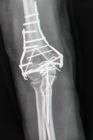
- ORIF distal humerus
- R/o Loose bodies
- Non-unions
Position
- Lateral
- Arm over radiolucent L bar
- Padding under axilla
Landmarks
- Olecranon process
Incision
- 10 cm longitudinal midline incision along posterior aspect of elbow
- Curve laterally around elbow to avoid ulnar nerve
Internervous Plane
- None, as extensor mechanism is detached with osteotomy
Superficial Dissection
- Deep fascia
- Identify ulnar nerve
- Mobilise ulnar nerve from its bed & hold it free with vascular tape
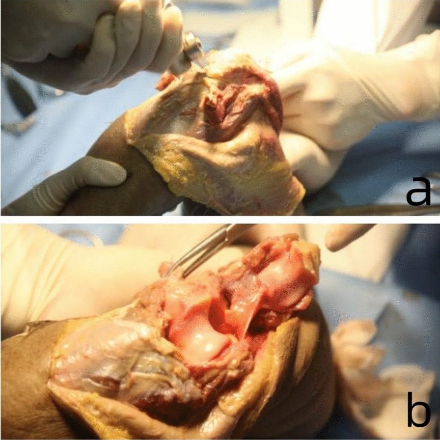
- Olecranon osteotomy
- Pre-drill & screw olecranon
- Score olecranon longitudinally
- V-shaped osteotomy about 2 cm from tip, apex directed distally
Deep Dissection
- Elevate triceps from back of humerus
- Subperiosteal dissection around medial & lateral borders of humerus as needed
- Transpose ulnar nerve anteriorly
Dangers
- Nerves
- Ulnar
- Radial
- Median
- Vessels
- Brachial artery
Reviewed by Professor Phong Tran, Head of Orthopaedic Surgery, Western Health. Last updated 10 October 2026.