
Position
- Supine
- Radiolucent arm board
- Exsanguinate & tourniquet
Landmarks
- Biceps tendon
- Mobile wad (BR, ECRL, ECRB)
- Styloid process of radius
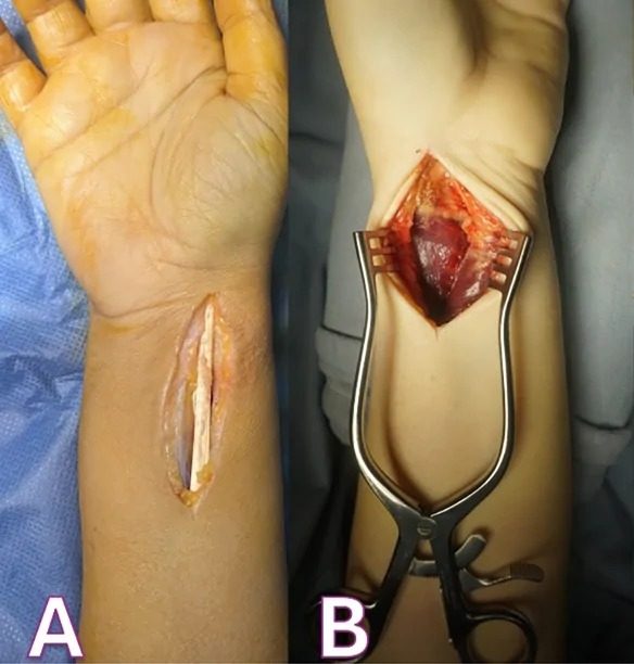
Incision
- Straight longitudinal incision
- Start: anterior flexor crease of elbow just lateral to biceps tendon
- End: Styloid process of radius
Internervous Plane
- Distally
- BR radial nerve
- FCR median nerve
- Proximally
- BR radial nerve
- PT median nerve
Superficial Dissection
- Fascia in line with skin incision
- Distally
- Identify & develop plane between BR & FCR
- Proximally
- Identify & develop plane between BR & PT
- (NB medial border of BR is surprisingly far across the forearm)
- Start distally & work proximally
- Identify superficial radial nerve (under BR)
- Proximally
- BR receives recurrent radial artery — ligate
- Retract BR & superficial radial nerve laterally
- Retract FCR & radial artery medially
Deep Dissection
- Proximal third
- Deepen the wound lateral to biceps tendon
- Biceps tendon inserts into bicipital tuberosity of radius
- Radial artery is superficial & medial to biceps tendon
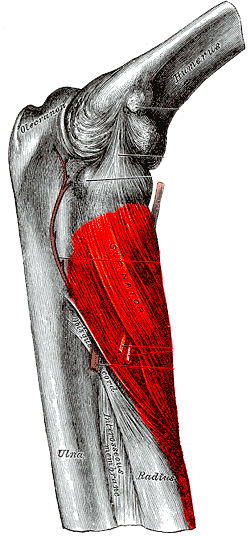
- Locate supinator
- Supinate forearm
- Displaces PIN laterally & posteriorly
- Incise supinator along the line of its broad insertion into anterior aspect of radius
- Continue subperiosteal dissection
- Avoid excessive retraction and don’t place retractors around radial neck
- PIN very very close
- Supinate forearm
- Deepen the wound lateral to biceps tendon
- Middle third
- Pronate forearm
- Exposes insertion of PT into lateral aspect of radius
- Detach insertion & retract PT medially
- Pronate forearm
- Distal third
- Detach PQ & FPL from attachments
Dangers
- Nerves
- PIN
- Superficial radial nerve
- Vessels
- Radial artery
- Recurrent radial arteries
Reviewed by Professor Phong Tran, Head of Orthopaedic Surgery, Western Health. Last updated 10 October 2026.