
Indications
Diagnostic vs Therapeutic
- Diagnostic
- Assess tears of TFCC
- Chondral damage to carpus
- Chronic wrist pain of unknown Diagnosis
- Therapeutic
- Distal radial & carpal Fracture
- Aid in ORIF distal radius fracture with mini-open procedure
- Percutaneous pinning of scaphoid
- Ligament tears
- Scapho-lunate instability
- Luno-triquetral instability
- Debride TFCC tears
- Synovectomy
- Removal of loose bodies
- Wrist lavage
- Arthrofibrosis
- Distal radial & carpal Fracture
Setup
- Tourniquet
- Traction
- essential for visualisation
- Traction tower & finger traps to 2 or 3 digits (usually IF & MF)
- If don’t have tower use shoulder holder & wt suspended off traps
- Overhead or traction device – so that elbow lies just off the table
- 10lb wt
- 2.7 mm scope
- essential
- 2.7mm or 2.9mm 30° or 70° scope
- 1.9mm scope also available for DRUJ, thumb CMCJ/ STTJ, MCPJ
- Infuse NS 1st
- Small joint instrumentation as well
Landmarks
- KEY IS KNOWLEDGE OF ANATOMY AND ACCURATE PORTAL PLACEMENT
- Draw portals, bony & tendon landmarks on skin prior to placement
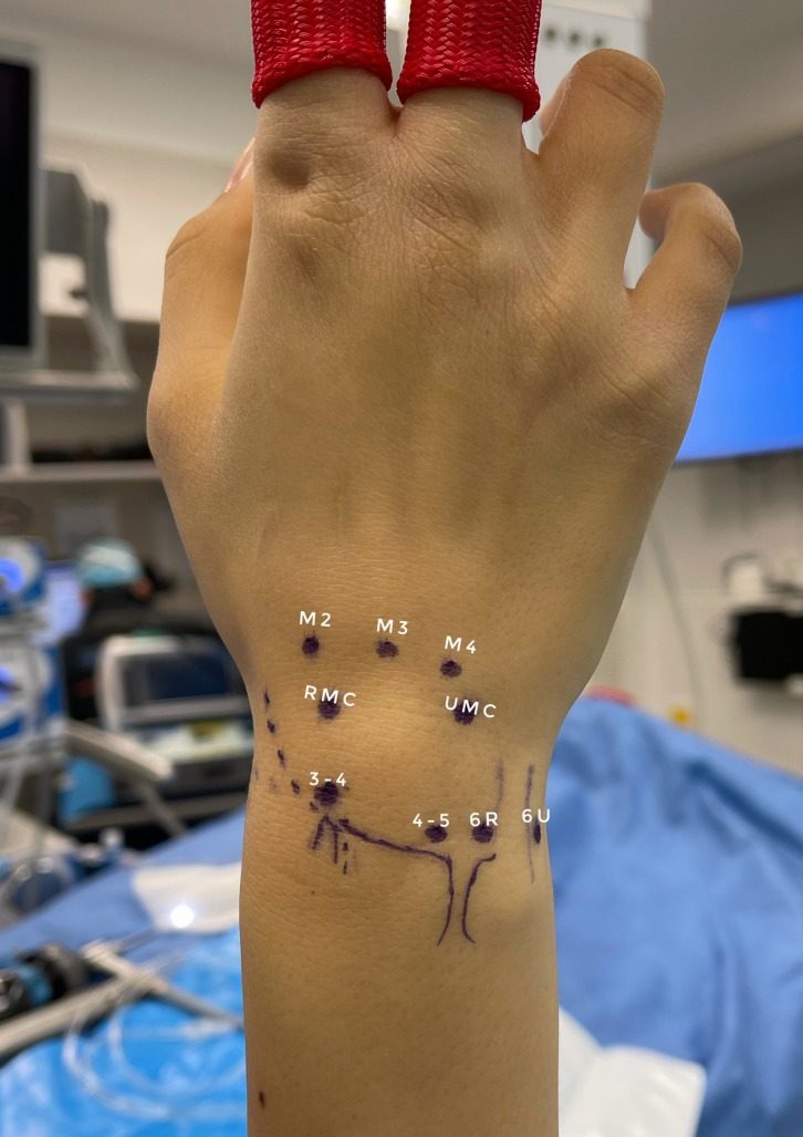
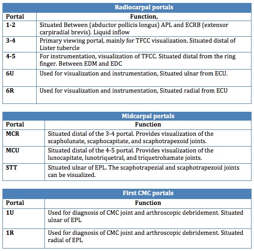
Portals
- Named according to interspace through which they pass
- Most common
- 3-4 portal
- 4-5 portal
- 6-R portal
- 6-U portal
- 3-4 Portal
- Feel Lister’s tubercle
- 1cm distal is soft spot between extensor compartments 3 & 4
- 4-5 Portal
- Roll finger over mobile 4th compartment to feel soft spot
- Is slightly proximal to 3-4 because of slope of radius
- 6-R & 6-U
- Named after their position about ECU
- Then
- Insufflate wrist with 3-5ml N/Saline into radiocarpal joint
- All portal incisions longitudinal to protect extensors
- Only incise skin
- Use blunt trocars to avoid radial n injury
- Pass trocar at 30-40° passing volar to conform to shape of radius
- Midcarpal Portals
- Two portals to view S-shaped midcarpal joint
- Made 1cm distal to 3-4 & 4-5 portals
- In soft spot between capitate & scaphoid
- 3-4 portal is primary viewing portal
- 4-5 or 6-R main working portal
- 6-U is inflow portal
- Outflow usually through IV tubing attached to scope
Diagnostic Procedure
- Start at radial styloid & scaphoid
- Work radial to ulnar
- Identify RadioScaphoCapitate Ligament & immediately beside is Long Radiolunate Ligament (extremely wide usually 3x width of RSCL)
- Next is Short RadioLunate Ligament (often see blood vessels along this ligament)
- Distal to short ligament is Scapholunate Lig
- Examine from membranous proximal portion to thicker dorsal ligamentous portion
- Follow ulnarly along lunate & its fossa to TFCC
- Articular disc should be taut like a trampoline (actual ballottement with probe should give same feeling = Trampoline test)
- Examine for tears » central or peripheral if not taut
- Don’t be caught out by ulnar styloid recess is normal finding at base of styloid not a tear
- Then to triquetrum
- Must probe both scapholunate joint & lunotriquetral joint for instability
- Next move to midcarpal joint with scope & probe using both midcarpal portals
- Most prominent feature is curve of head of capitate
- Reverse look & view scapholunate joint & lunotriquetral joint
- STTJ can be seen by passing completely radially
Carpal Instability
- Must look from radiocarpal & midcarpal joints
- Both joint ligaments should be tight & concave
- Can do “poor man’s arthrogram”
- Ie inflow in RCJ outflow in MCJ
- If flow then have tear in ligament
Arthroscopic Classification
| Grade | Description | Treatment |
|---|---|---|
| I | Attenuation or haemorrhage within ligament. No step | Cast immobilisation |
| II | Incongruency or step-off in midcarpal space | Arthroscopic pinning. Use K wire as joystick to reduce |
| III | Step-off on both sides. Probe may be passed between bones | Arthroscopic or open repair |
| IV | Gross instability | Open repair |
TFCC Injuries
- Use 4-5 portal as visual portal & 6-R as working portal
- Degenerative or Traumatic
- Central or Peripheral
- With or without DRUJ instability
- Radial or Ulnar avulsions
- ± Styloid Fracture
- Debride central tears acute or degenerative
- Attempt repair of peripheral tears
- If unstable DRUJ have to reinforce DRUL or PRUL with strip of ECU
- If degenerative tear & ulnar plus need to add ulnar shortening to debridement
Distal Radial Fractures
- Critical tolerance for articular incongruity = 2mm
- Recent evidence now says 1mm
- A/scopic assisted helps to achieve congruity
- Also now are seeing high incidence of scapholunate & lunotriquetral ligament injuries
- Technique
- Usually wait 2-7/7 for bleeding
- Usual set up
- 3-4 visual portal, 4-5 or 6-R as working portals,6-U as inflow
- Occasionally use 1-2 portal
- Use K-wires placed under I-I control to joystick fragments as well as small pick etc through working portal
- Wire fixation as per usual once reduction obtained
- If see die punch have to open
Reviewed by Professor Phong Tran, Head of Orthopaedic Surgery, Western Health. Last updated 10 October 2026.