
Definition
- Obsolete term for a tumour with branching staghorn vessels
- WHO now places almost all cases within solitary fibrous tumour (SFT)
- Intermediate, rarely metastasising, with a malignant subset
Epidemiology
- Adults, most often middle aged, with no strong sex predilection
- Pleura, meninges, deep thigh, pelvis, retroperitoneum and head and neck
- Primary bone involvement is rare
- Meningeal haemangiopericytoma is now termed meningeal SFT
Pathology
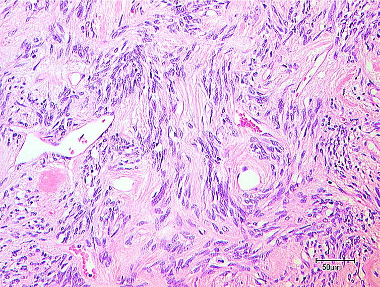
- Patternless ovoid to spindle cells in variably collagenous stroma
- Thin walled, branching, dilated staghorn vessels
- NAB2 to STAT6 fusion on chromosome 12
- Strong nuclear STAT6 staining is the diagnostic marker
- CD34 usually positive
- Haemangiopericytoma-like vessels also occur in synovial sarcoma and mesenchymal chondrosarcoma
Clinical Features
- Slowly enlarging, usually painless deep mass
- Pressure symptoms from large pelvic or retroperitoneal tumours
- Doege-Potter syndrome, paraneoplastic hypoglycaemia from tumour IGF-2 secretion in large tumours
- Bone lesions present with pain or pathological fracture
Investigations
- MRI shows a well circumscribed, heterogeneous, avidly enhancing mass with flow voids
- CT angiography shows hypervascularity
- Core biopsy at a sarcoma centre with STAT6 immunohistochemistry
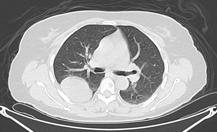
- Staging CT chest, with liver and bone other metastatic sites
Classification
- Histology alone does not reliably predict behaviour
- Risk models combine age, tumour size, mitotic rate and necrosis
- Tumours graded low, intermediate or high risk for metastasis
Management
- Wide surgical excision is the primary treatment
- Preoperative embolisation reduces blood loss in vascular tumours
- Radiotherapy for close or positive margins and unresectable disease
- Conventional chemotherapy has modest activity
- Pazopanib, or temozolomide with bevacizumab, for advanced disease
- Hypoglycaemia resolves after complete resection
Prognosis
- Most behave indolently after complete excision
- Recurrence and metastasis may occur beyond ten years
- Long term follow up, particularly for intermediate and high risk tumours
Reviewed by Professor Phong Tran, Head of Orthopaedic Surgery, Western Health. Last updated 10 October 2026.