
Definition
- Condition at the benign end of the spectrum of histiocytosis X associated with unifocal or multifocal granulomas containing Langerhans cells & eosinophils
Terminology
- Histiocytosis X comprises a spectrum of disorders.
- EG
- most benign.
- Hand-Schuller-Christian disease
- exophthalmos, diabetes insipidus, & bone destruction
- Letterer-Siwe syndrome
- Most severe & potentially fatal
- characterised by hepatosplenomegaly, lymphadenopathy, anaemia, acute infections & a downhill course
- EG
Epidemiology
- First two decades of life
- Slightly more common in males
Pathology
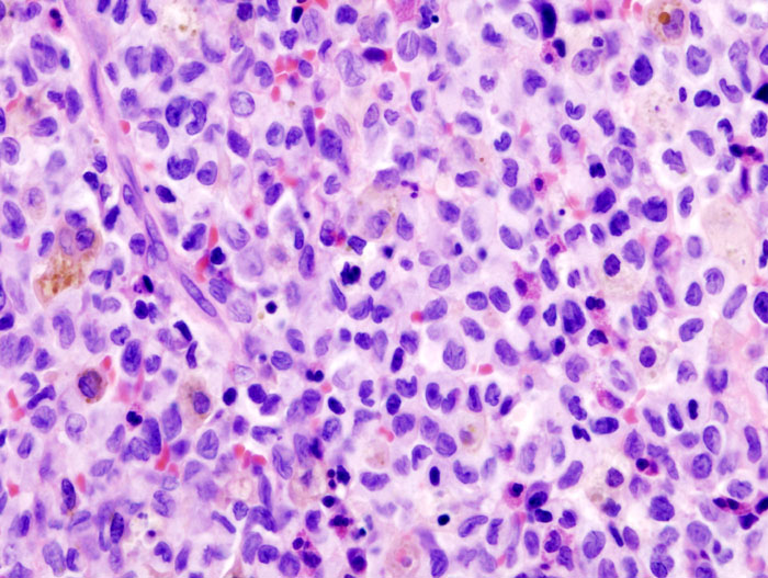
- cell of origin is the Langerhans cell, a dendritic antigen presenting cell that is found all throughout the body, particularly in the skin & bones
- Langerhans cell contains Birbeck granules, & has a nucleus with a deep cleft
- Other cells found in the granulomas include eosinophils, PMNLs, giant cells, & mononuclear cells. The Langerhans cells are arranged in clusters.
- Necrosis is very commonly seen
- Grossly the lesion is soft, tan to reddish & occasionally greenish
Distribution
- Skull, spine, pelvis & long bones, particularly the femur
- Most commonly unifocal but may be multifocal – up to 50%
- Has a preference for the skull & femur in children (sites of haematopoietic tissue) & the pelvis, ribs & skull in adults
Clinical
- Commonest symptom is pain. The pain is often worse at night
- May be accompanied by a palpable mass, fever & a limited range of motion
- Skin lesions are often seen
Investigations
Laboratory
- Rarely helpful. Occasional leukocytosis & elevated ESR. Usually no eosinophilia
X-ray
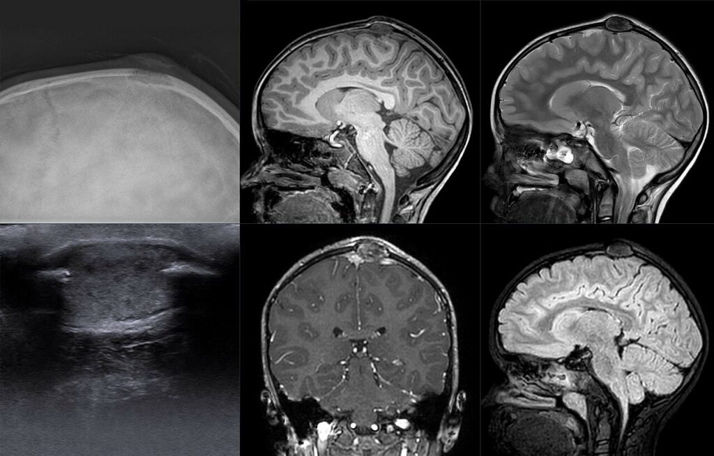
- XR typically shows lucent lesion sometimes with sclerosis.
- lesion is typically in the medullary canal of the diaphysis of a long bone
- It can occur in the epiphysis, however, & cross open growth plates
- It may cause cortical destruction
- It is the great mimicker & is in the differential diagnosis of any skeletal lesion. There may be expansion or periosteal reaction
- In the spine causes vertebra plana
MRI
- typically low T1 signal & high T2 signal. Surrounding oedema
Differential Diagnosis
- Osteomyelitis
- Ewing’s sarcoma
Treatment
- For EG, treatment is usually observation & the tendency is for resolution. Healing usually takes at least 5 months.
- If there is ongoing pain or a lesion in a weight bearing bone then intralesional corticosteroids or curettage & bone grafting is used
- Radiotherapy can be used for inaccessible lesions
Reviewed by Professor Phong Tran, Head of Orthopaedic Surgery, Western Health. Last updated 10 October 2026.