
Definition
- Benign bone forming tumour histologically similar to osteoid osteoma but over 2 cm
- About 1% of primary bone tumours
- Can behave aggressively, with an epithelioid variant that may mimic osteosarcoma
Epidemiology
- Age 10 to 30 years
- Male predominance, about 2 to 1
- About 40% arise in the posterior elements of the spine
- Long bones are next most common, often diaphyseal or metaphyseal
- Rare in the small bones of hands and feet
Clinical Features
- Dull pain, less night pain and less NSAID response than osteoid osteoma
- Painful scoliosis and nerve root symptoms in spinal lesions
- Swelling and tenderness in superficial sites
- Neurological deficit from spinal canal extension
Investigations
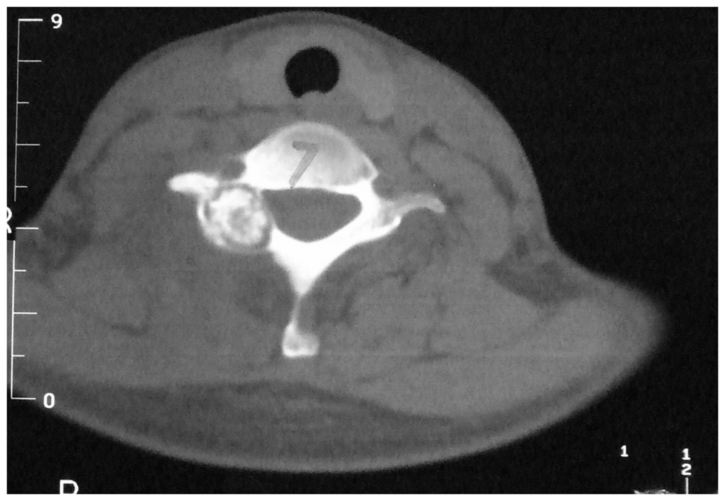
- Expansile lytic lesion with central mineralisation
- CT defines cortical breach and spinal extent
- Biopsy to exclude osteosarcoma
- Bone scan shows intense uptake
- MRI shows surrounding oedema and soft tissue extension
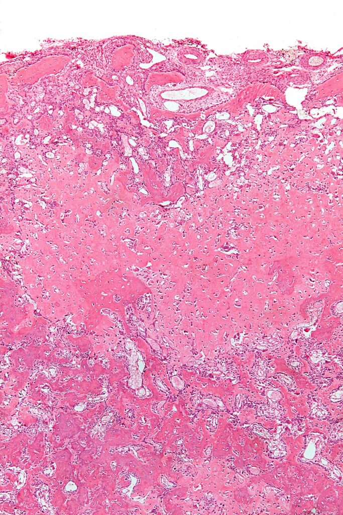
- Histology shows osteoid and woven bone rimmed by osteoblasts, without the permeative growth of osteosarcoma
Management
- Extended curettage with adjuvant and bone grafting, or en bloc excision
- Recurrence up to about 20% after curettage
- Stabilise the spine if resection causes instability
- Radiofrequency ablation is less suitable than for osteoid osteoma because of size
- Embolisation before surgery for vascular spinal lesions
Differential Diagnosis
- Osteoid osteoma
- Aneurysmal bone cyst
- Osteosarcoma
Reviewed by Professor Phong Tran, Head of Orthopaedic Surgery, Western Health. Last updated 10 October 2026.