
Definition
- Separate ossicle above a hypoplastic dens, with a gap between it and the C2 body
Aetiology
- Evidence suggests an unrecognised and untreated odontoid fracture
- Congenital origin is possible in some
- Associated with Down syndrome, Morquio syndrome and Klippel-Feil syndrome
Classification
| Type | Description |
|---|---|
| Orthotopic | Ossicle moves with the anterior arch of C1 |
| Dystopic | Ossicle fused to the clivus or displaced |
Clinical Features
- Incidental finding in some
- Neck pain and torticollis
- Myelopathy or transient neurology after minor trauma
- Vertebrobasilar ischaemia
Investigations
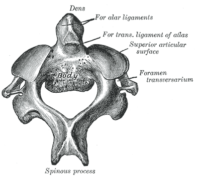
- Open mouth and lateral radiographs
- Flexion and extension radiographs for atlantoaxial instability
- CT shows smooth corticated margins, unlike an acute fracture
- MRI for cord compression and signal change
Differential Diagnosis
- Acute odontoid fracture
- Persistent ossiculum terminale
Management
- Asymptomatic stable cases observed with activity counselling
- Instability on flexion and extension X-rays, consider C1 to C2 fusion
- Neurological deficit or myelopathy, fusion
- Posterior C1 to C2 fusion with transarticular or C1 lateral mass and C2 screws
Complications
- Sudden cord injury with minor trauma in unstable cases
- Nonunion of fusion
- Vertebral artery injury during screw placement
Reviewed by Professor Phong Tran, Head of Orthopaedic Surgery, Western Health. Last updated 10 October 2026.