
Epidemiology
- Most frequent physeal injury in children
- About 80% are Salter-Harris I or II
Aetiology
- Fall on outstretched hand
- Repetitive loading in gymnasts causes distal radial physeal stress injury
Clinical Features
- Wrist swelling and deformity
- Check median nerve function
- Check skin for puncture wounds suggesting an open fracture
Investigations
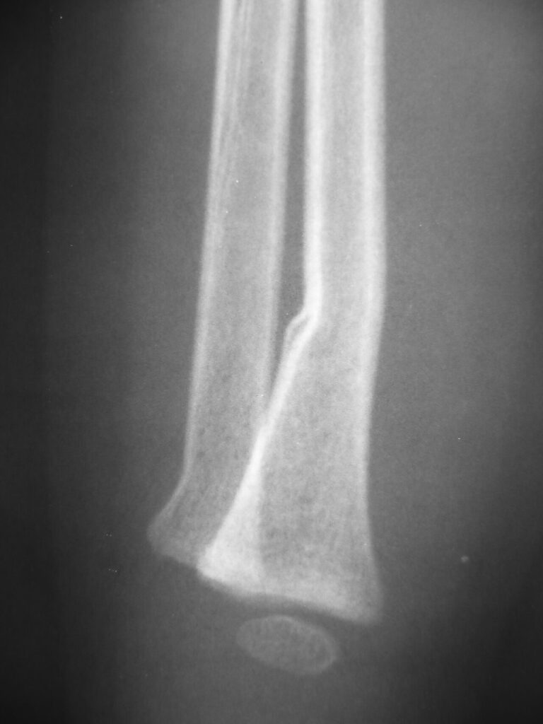
- AP and lateral wrist radiographs
- Look for associated ulnar styloid or distal ulnar physeal injury
Management
- Closed reduction and above elbow cast for displaced fractures
- Gentle reduction under anaesthesia to protect the physis
- Remodelling potential is high in young children
- Accept residual angulation in children with substantial growth left
- Do not remanipulate after about 7 days as this risks growth disturbance
- Percutaneous smooth K wires for unstable fractures or swelling
- ORIF for irreducible, intra-articular type III and IV or open fractures
Complications
- Growth arrest in a small minority
- Ulnar positive variance after radial arrest
- Median nerve compression and acute carpal tunnel syndrome
- Loss of reduction
Prognosis
- Good outcome in most
- Follow up radiographs for growth disturbance after higher energy injuries
- Growth arrest may present late with wrist pain and ulnar impaction
Reviewed by Professor Phong Tran, Head of Orthopaedic Surgery, Western Health. Last updated 10 October 2026.